Introduction
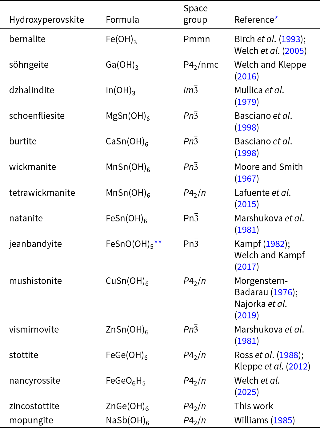
Hydroxyperovskites are frameworks of corner-linked octahedra in which all oxygen atoms form hydroxyl groups and each is a donor and acceptor. The cavity A-site of normal perovskites is empty in hydroxyperovskites. Coupled with moderately strong hydrogen bonding, the absence of an A cation results in a framework of highly tilted octahedra. Two stoichiometries have been recognised so far, corresponding to single and double hydroxyperovskites, denoted B(OH)3 and BB′(OH)6, respectively, where B and B′ are metal cations. Double hydroxides are combinations of divalent and tetravalent cations, and are predominantly stannates, although a germanate and a silicate (synthetic) are also known. The single exception to the B 2+/B′4+ combination is mopungite NaSb(OH)6. The crystal chemistry of hydroxyperovskites is reviewed by Mitchell et al. (Reference Mitchell, Welch and Chakmouradian2017). Herein, we describe zincostottite, the second natural germanate hydroxyperovskite. Including the recently approved nancyrossite, there are now 15 natural hydroxyperovskites (Table 1).
Natural hydroxyperovskites

* Structure.
** Formula reported by Welch and Kampf (Reference Welch and Kampf2017).
Zincostottite is named as the Zn analogue of stottite, Fe2+Ge(OH)6. The new mineral and the name have been approved by the Commission on New Minerals, Nomenclature and Classification of the International Mineralogical Association (IMA2024-024, Kampf et al., Reference Kampf, Désor, Ma, Welch and Möhn2024; Warr symbol: Zsto). The description is based upon one holotype specimen and one cotype specimen deposited in the collections of the Natural History Museum of Los Angeles County, 900 Exposition Boulevard, Los Angeles, CA 90007, USA, catalogue numbers 76278 and 76279, respectively.
Occurrence
Zincostottite was found on specimens from the Tsumeb mine, Tsumeb, Namibia (19°15′S, 17°42′E). An overview of the mineralogy and geology of the Tsumeb deposit can be found in Gebhard (Reference Gebhard1999) and von Bezing (Reference von Bezing2007). Zincostottite occurs on fracture surfaces in ore containing a mixture of germanite, chalcocite, bornite and tennantite-(Zn). Other minerals found in association with zincostottite are siderite, malachite and quartz. The specimens come from the collection of Karl Seifert, who was a mining engineer at Tsumeb from 1963 to 1967. The 2nd oxidation zone of the deposit was being mined during that period. Zincostottite probably originates from the Ge-rich horizon at Level 30 of the mine, ∼900 m below the surface and near the top of the 2nd oxidation zone. Zincostottite is a secondary oxidation-zone mineral.
Physical and optical properties
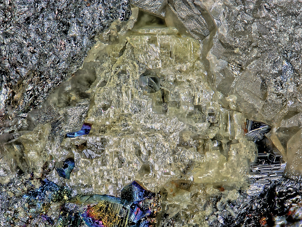
Zincostottite occurs as heavily etched remnants of equant or tabular crystals, up to ∼1 mm in diameter (Fig. 1). No crystal forms could be measured and no twinning was observed. The mineral is transparent and has a white streak. The mineral does not fluoresce in long- or short-wave ultraviolet light. The Mohs hardness is ∼4.5 based on scratch tests. Crystals are brittle with irregular stepped fracture. Cleavage is good on {100} and poor on {001}. The density could not be measured because crystals exceed the density of available density fluids. The calculated density is 3.834 g·cm–3 for the empirical formula using the single-crystal cell. At room temperature, the mineral is soluble in dilute HCl.
Heavily etched zincostottite crystal with chalcocite crystals in vug lined with siderite crystals on cotype specimen (#76278); FOV 1.13 mm across.

Optically, zincostottite is uniaxial (–) with indices of refraction ω = 1.785(5) and ε = 1.765(5) measured in white light. The mineral is nonpleochroic. The Gladstone–Dale compatibility index (Mandarino, Reference Mandarino2007) is 0.051 for the empirical formula in the range of good compatibility.
Raman spectroscopy
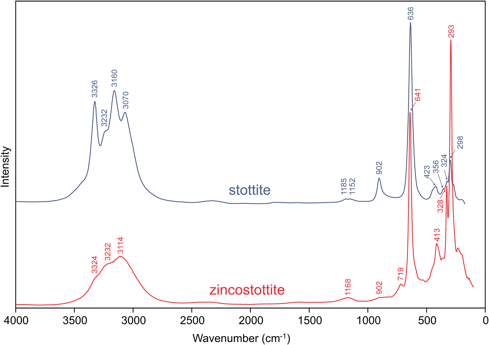
Raman spectroscopy was done on a Horiba XploRA PLUS using a 532 nm diode laser, 100 μm slit, 1800 grooves/mm diffraction grating and a 100× (0.9 NA) objective. The spectrum from 4000 to 100 cm–1 is compared with that of stottite (RRUFF #R120089) in Fig. 2. The spectra are seen to be very similar. The (OH)-stretching bands in the 3400 to 3000 cm–1 range are much better resolved in the stottite spectrum. Earlier studies have not attempted to assign specific framework modes to the bands at lower wavenumbers (Kleppe et al., Reference Kleppe, Welch, Crichton and Jephcoat2012).
Raman spectra of stottite and zincostottite.

Composition
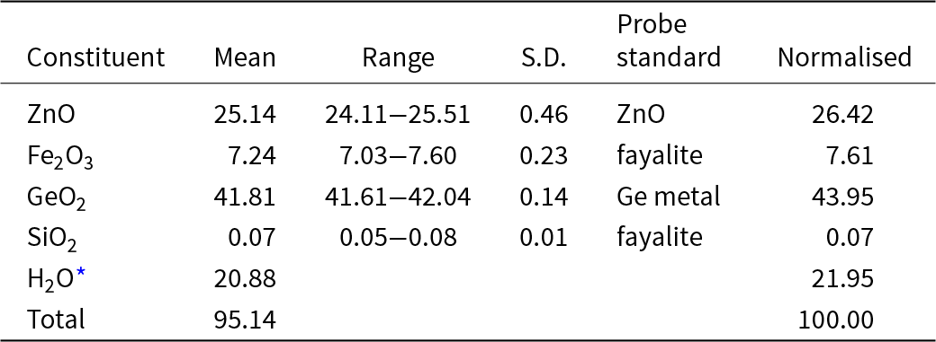
Electron probe microanalyses (EPMA) were done at Caltech on a JXA-iHP200F electron microprobe in WDS mode. The analytical conditions were 15 kV accelerating voltage, 5 nA beam current and 10 μm beam diameter. Analyses fell into two groups, one with almost equal amounts of Zn and Fe and the other with Zn:Fe ≈ 3:1. No zonation of Zn and Fe was observed in the crystals analysed. Below we report the analyses with Zn ≫ Fe, which are also consistent with the crystal structure refinement reported below. Insufficient material is available for the determination of H2O, so it is calculated based on the structure (O = 6 and Ge+Si = 1). The crystal structure refinement and OccQP analysis (Wright et al., Reference Wright, Foley and Hughes2000) indicate that all Fe in the structure crystal is in the ferric state (and structure refinements on several other zincostottite crystals are also consistent with all Fe being in the ferric state). Consequently, for the EPMA, all Fe has been calculated as +3. Nevertheless, we acknowledge that the presence of some Fe2+ is possible in some crystals of zincostottite, however because Fe is not an essential element, its oxidation state is incidental to the definition of the species. There is not enough material for Mössbauer spectroscopy.
The crystals did not take a good polish. The low analytical total is attributed to the somewhat irregular surface. Analytical data are given in Table 2. The empirical formula based on O = 6 atoms per formula unit (apfu) is (Zn0.77Fe3+0.23)Σ1.00Ge1.00O6H5.77. The ideal formula is ZnGe(OH)6, which requires ZnO 33.91, GeO2 43.58, H2O 22.52, total 100 wt.%.
Compositional data in wt.% for zincostottite

* Based on the structure (O = 6 apfu).
X-ray crystallography and structure refinement
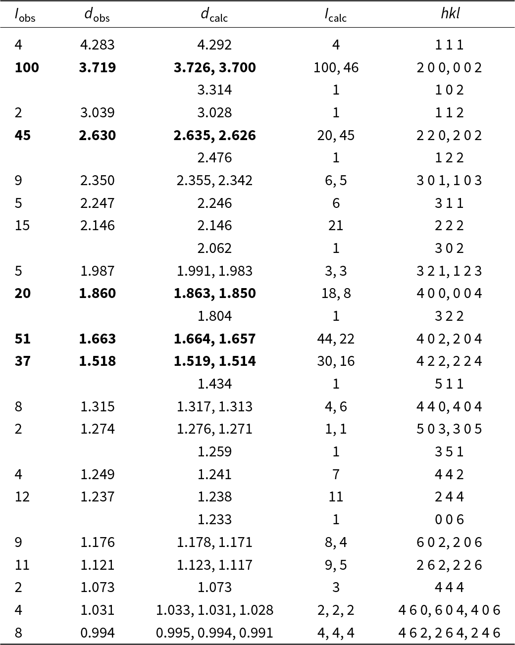
Powder X-ray diffraction data were recorded using a Rigaku R-Axis Rapid II curved imaging plate microdiffractometer with monochromatised MoKα radiation. A Gandolfi-like motion on the φ and ω axes was used to randomise the sample. Observed d-values and intensities were derived by profile fitting using JADE Pro software (Materials Data, Inc.). The powder data are presented in Table 3. The unit-cell parameters refined from the powder data using JADE Pro with whole-pattern fitting (space group P42/n) are a = 7.452(3), c = 7.400(4) Å and V = 411.0(4) Å3.

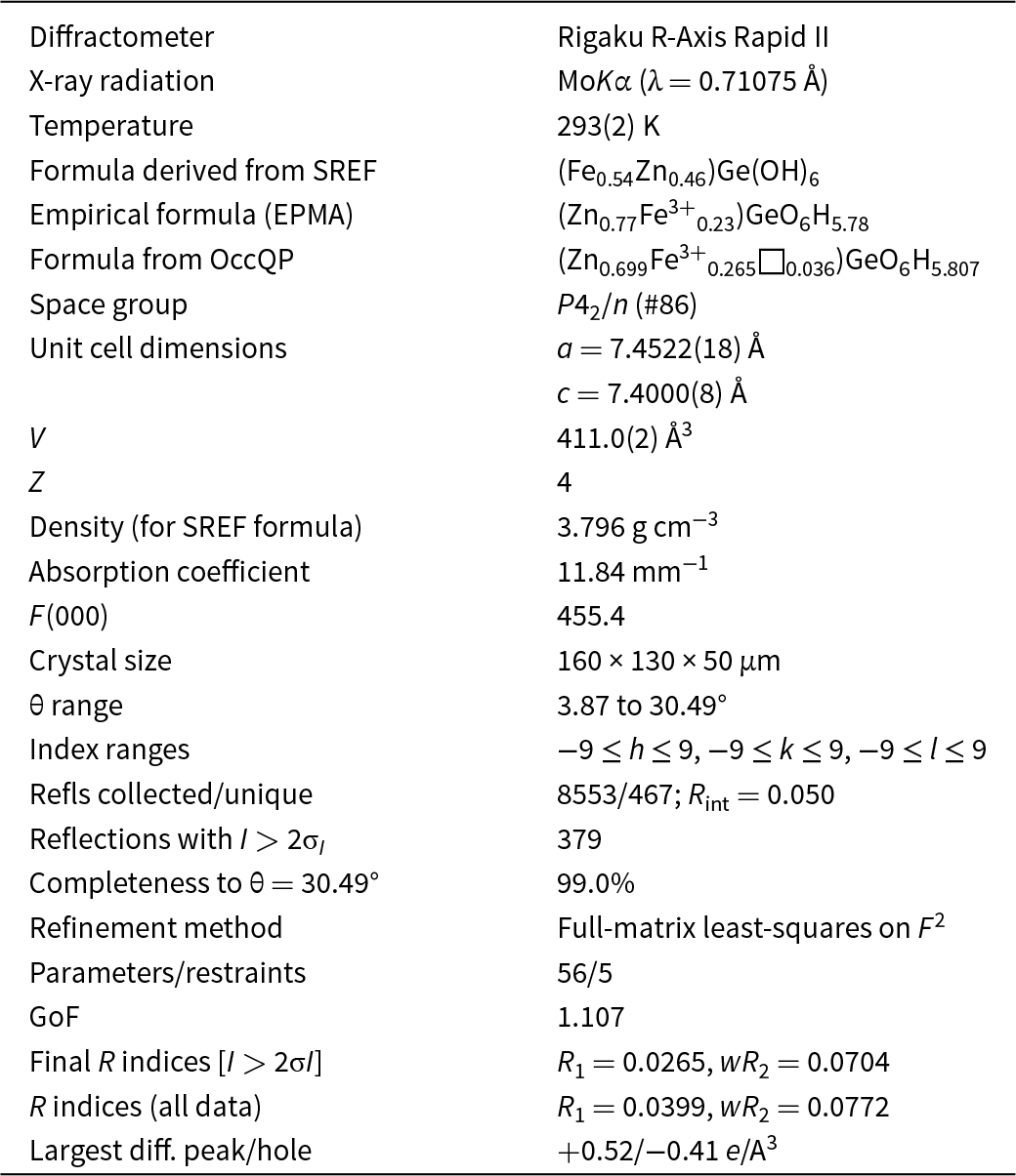
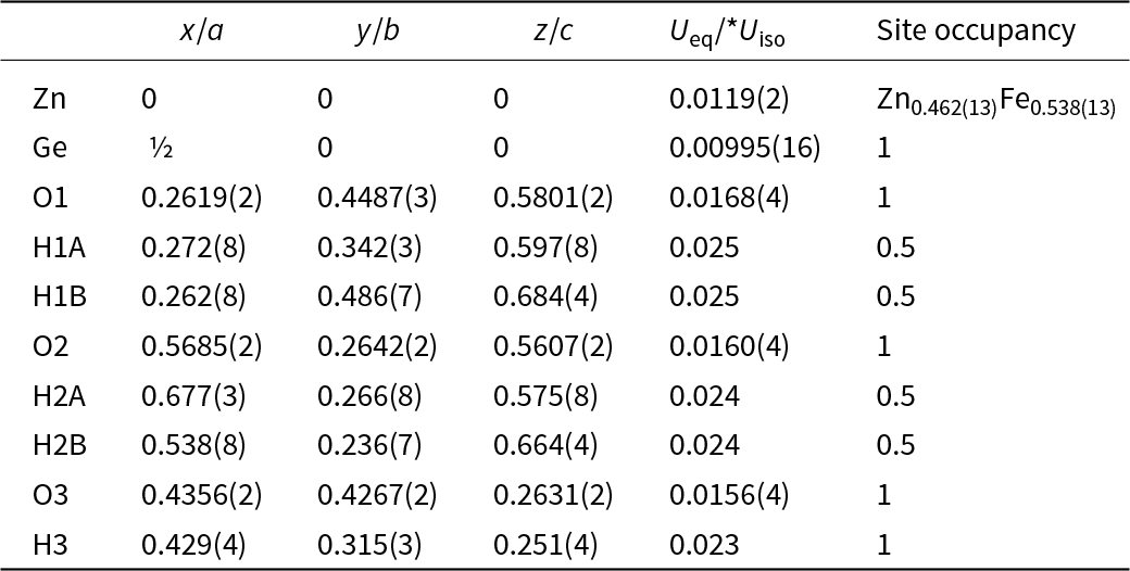
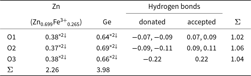
Single-crystal X-ray studies were done on the same diffractometer and radiation noted above. The Rigaku CrystalClear software package was used for processing the structure data, including the application of an empirical absorption correction using the multi-scan method with ABSCOR (Higashi, Reference Higashi2001). The structure was solved using SHELXT (Sheldrick, Reference Sheldrick2015a). Neutral atomic scattering factors were used (Wilson, Reference Wilson1992). Refinement proceeded by full-matrix least-squares on F 2 using SHELXL-2016 (Sheldrick, Reference Sheldrick2015b). The Zn site was refined with joint occupancy by Zn and Fe resulting in Zn0.462(13)Fe0.538(13) for a site-scattering value of 111.39±1.45 e per unit cell. This compares to a site-scattering value of 116.32 e per unit cell based on the EPMA (Zn0.77Fe0.23). It is worth noting that the structure crystal was checked by EDS following data collection and confirmed to be consistent with the EPMA composition. A possible explanation for the difference is that there is a small vacancy at the site. To test this as well as to confirm the oxidation state of Fe, we analysed the site using OccQP (Wright et al., Reference Wright, Foley and Hughes2000), a program that uses quadratic equations in a constrained least-squares formulation to optimise occupancy assignments based upon site scattering, chemical composition, charge balance, bond valence and cation-anion bond lengths. With the composition allowed to vary freely among the cations Zn, Fe2+ and Fe3+, the optimal site occupancy calculated with OccQP is Zn0.699Fe3+0.265Fe2+0.000□0.036. This occupancy fits the site scattering and cation site valence perfectly. The OccQP occupancy also provides a reasonable fit with the EPMA and confirms all Fe to be +3. The formula based on the OccQP analysis of the Zn site is (Zn0.699Fe3+0.265□0.036)GeO6H5.807. We note that the new Fe3+-dominant stottite-subgroup mineral nancyrossite (Welch et al., Reference Welch, Najorka, Kleppe, Kampf and Spratt2025), Fe3+GeO6H5, was found in the same Ge-rich horizon at Level 30 of the Tsumeb mine from which the zincostottite specimens are presumed to have come.
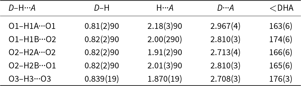
Difference-Fourier synthesis revealed the likely H sites. The H sites were refined with a soft restraint of 0.82(2) Å on the O–H distances and with the U eq set to 1.5 times that of the associated O atoms. The highly directional O–H···O linkages in hydroxyperovskites allow plausible H atoms in the list of difference-Fourier maxima to be recognised (Mitchell et al., Reference Mitchell, Welch and Chakmouradian2017). Furthermore, it is known that some H sites in hydroxyperovskites are half-occupied (Basciano et al., Reference Basciano, Peterson, Roeder and Swainson1998; Lafuente et al., Reference Lafuente, Yang and Downs2015; Welch and Kleppe, Reference Welch and Kleppe2016; Welch et al., Reference Welch, Najorka and Wunder2024). Hydroxyperovskites with space group P42/n, such as stottite, have one fully occupied and four half occupied H (Lafuente et al., Reference Lafuente, Yang and Downs2015; Welch and Kleppe, Reference Welch and Kleppe2016). Zincostottite has one fully occupied and four half occupied H sites, as is characteristic of P42/n hydroxyperovskites. No evidence of merohedral twinning (0 1 0/1 0 0/0 0  $\bar 1$) was found in difference-Fourier maps. Also, no superlattice reflections that would indicate ordering of Zn and Fe3+ were seen in the diffraction patterns of any of the crystals examined, including those which had nearly equal amounts of Zn and Fe3+.
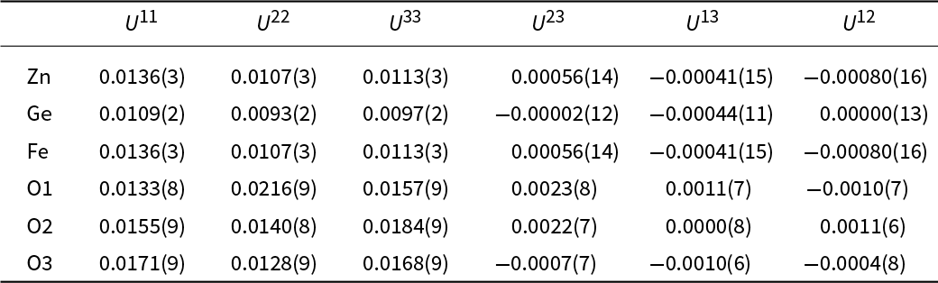
Data collection and refinement details are given in Table 4, atom coordinates and displacement parameters in Tables 5 and 6, selected bond distances and angles in Tables 7 and 8 and a bond valence analysis in Table 9. Note that the Zn-site occupancy indicated by OccQP (Zn0.699Fe3+0.265□0.036) was used for the bond-valence calculations. The crystallographic information file has been deposited with the Principal Editor of Mineralogical Magazine and is available as Supplementary material (see below).
Data collection and structure refinement details for zincostottite

Notes: R int = Σ|F o2–F o2(mean)|/Σ[F o2]. GoF = S = {Σ[w(F o2–F c2)2]/(n–p)}1/2. R 1 = Σ||F o|–|F c||/Σ|F o|. wR 2 = {Σ[w(F o2–F c2)2]/Σ[w(F o2)2]}1/2; w = 1/[σ2(F o2) + (aP)2 + bP] where a is 0.032, b is 0.25 and P is [2F c2 + Max(F o2,0)]/3.
Atom positions and site occupancies for zincostottite

Displacement parameters (Å)2 for zincostottite

Selected bond lengths (Å) for zincostottite

Hydrogen bonds for zincostottite

Bond valences analysis for zincostottite. Values are in valence units (vu)*

* Bond-valence parameters are from Gagné and Hawthorne (Reference Gagné and F.C2015). Hydrogen-bond valences are based on O–O bond lengths from Ferraris and Ivaldi (Reference Ferraris and Ivaldi1988). Negative values indicate donated hydrogen-bond valence.
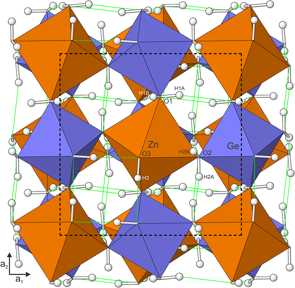
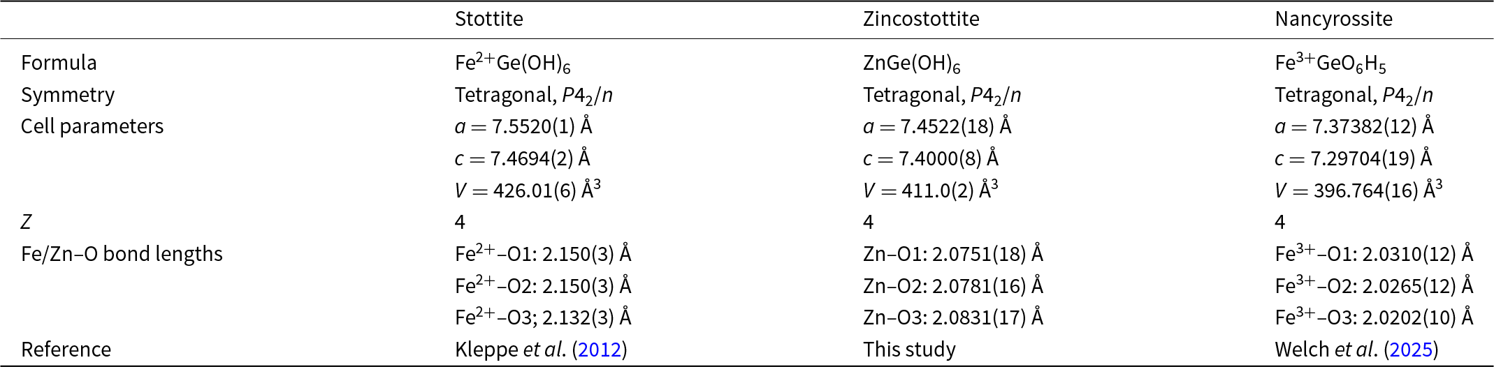
Zincostottite is isostructural with stottite (Ross et al., Reference Ross, Bernstein and Waychunas1988) and other double hydroxyperovskites in the stottite subgroup (Mitchell et al., Reference Mitchell, Welch and Chakmouradian2017). The structure is a 3D framework consisting of alternating Zn(OH)6 and Ge(OH)6 octahedra sharing OH corners (Fig. 3). The hydrogen bonding scheme is the same as that delineated by Ross et al. (Reference Ross, Chaplin and Welch2002) based on O–O bond distances and later confirmed by Kleppe et al. (Reference Kleppe, Welch, Crichton and Jephcoat2012) with the determination of H atom positions. Selected data for stottite, zincostottite and nancyrossite are compared in Table 10.
The zincostottite structure viewed down [001]. Hydrogen bonds are shown as light green lines. The unit cell outline is shown with dashed lines. The figure was created using ATOMS, version 6.5 (Shape Software, Kingsport, Tennessee, USA).

Selected data for stottite and zincostottite.

Discussion
Hydroxyperovskites have been recognised as having potential as materials for use in photocatalysis and electrolytic catalysis (Evans et al., Reference Evans, Wu, Seshadri and Cheetham2020). Minerals such as jeanbandyite, FeSnO6H5, and nancyrossite, FeGeO6H5, have proton deficiencies (five instead of six H pfu) and may have novel H behaviour, such as high proton mobility. Proton conductivity in these materials and related hydroxyperovskites is unexplored. Hydroxyperovskites, in general, are an overlooked group of structures that deserve further attention as potential functional materials.
Supplementary material
The supplementary material for this article can be found at https://doi.org/10.1180/mgm.2024.81.
Acknowledgements
Two anonymous reviewers and Structures Editor Peter Leverett are thanked for their constructive comments on the manuscript. The EPMA was carried out at the Caltech GPS Division Analytical Facility, which is supported, in part, by NSF Grant EAR-2117942. A portion of this study was funded by the John Jago Trelawney Endowment to the Mineral Sciences Department of the Natural History Museum of Los Angeles County.
Competing interests
The authors declare none.