Introduction
Major depressive disorder (MDD) is a highly prevalent and disabling mental disorder with significant morbidity, mortality, and cost (Bromet et al., Reference Bromet, Andrade, Hwang, Sampson, Alonso, de Girolamo and Kessler2011; Greenberg et al., Reference Greenberg, Fournier, Sisitsky, Simes, Berman, Koenigsberg and Kessler2021; Kessler & Bromet, Reference Kessler and Bromet2013; Whiteford et al., Reference Whiteford, Degenhardt, Rehm, Baxter, Ferrari, Erskine and Vos2013). The World Health Organization projected that MDD will be the leading cause of global disease burden by 2030 (World Health Organization, 2008). Although substantial efforts have been made in the past decades, the etiology of MDD remains elusive. Advances in neuroimaging techniques have made it increasingly feasible to investigate the neuropathology of MDD. Benefiting from the ability to precisely map function to underlying neuroanatomy, functional neuroimaging is crucial for studying brain function in health and disease. Cerebral blood flow (CBF), normally coupled to brain metabolism and neuronal activity, is one of the most frequently used and physiologically relevant functional measures (Buxton, Reference Buxton2021; Lecrux, Bourourou, & Hamel, Reference Lecrux, Bourourou and Hamel2019). Positron emission tomography (PET) and single photon emission computed tomography (SPECT) have traditionally been adopted to measure CBF (Wintermark et al., Reference Wintermark, Sesay, Barbier, Borbély, Dillon, Eastwood and Yonas2005). Arterial spin labeling (ASL), a noninvasive magnetic resonance imaging (MRI) technique, can rapidly quantify CBF using magnetically labeled arterial blood water as an endogenous tracer (Haller et al., Reference Haller, Zaharchuk, Thomas, Lovblad, Barkhof and Golay2016; Hernandez-Garcia, Lahiri, & Schollenberger, Reference Hernandez-Garcia, Lahiri and Schollenberger2019). With use of these techniques, numerous studies have shown CBF changes in MDD patients (Bench et al., Reference Bench, Friston, Brown, Scott, Frackowiak and Dolan1992; Cantisani et al., Reference Cantisani, Stegmayer, Bracht, Federspiel, Wiest, Horn and Walther2016; Chen et al., Reference Chen, Bian, Jiang, Cui, Ji, Liu and Zhuo2016; Cooper et al., Reference Cooper, Chin Fatt, Liu, Grannemann, Carmody, Almeida and Trivedi2020; Duhameau et al., Reference Duhameau, Ferre, Jannin, Gauvrit, Verin, Millet and Drapier2010; Ho et al., Reference Ho, Wu, Shin, Liu, Tapert, Yang and Yang2013; Jarnum et al., Reference Jarnum, Eskildsen, Steffensen, Lundbye-Christensen, Simonsen, Thomsen and Larsson2011; Kaichi et al., Reference Kaichi, Okada, Takamura, Toki, Akiyama, Higaki and Awai2016; Krausz et al., Reference Krausz, Freedman, Lester, Barkai, Levin, Bocher and Bonne2007; Li et al., Reference Li, Yang, Zhu, Zhou, Han, Yin and Chen2018; Lui et al., Reference Lui, Parkes, Huang, Zou, Chan, Yang and Gong2009; Monkul et al., Reference Monkul, Silva, Narayana, Peluso, Zamarripa, Nery and Soares2012; Ota et al., Reference Ota, Noda, Sato, Hattori, Teraishi, Hori and Kunugi2014; Périco et al., Reference Périco, Skaf, Yamada, Duran, Buchpiguel, Castro and Busatto2005; Sahib et al., Reference Sahib, Loureiro, Vasavada, Kubicki, Joshi, Wang and Narr2020; Savitz et al., Reference Savitz, Nugent, Cannon, Carlson, Davis, Neumeister and Drevets2012; Vardi et al., Reference Vardi, Freedman, Lester, Gomori, Chisin, Lerer and Bonne2011; Vasic et al., Reference Vasic, Wolf, Gron, Sosic-Vasic, Connemann, Sambataro and Wolf2015), establishing CBF as a potential imaging biomarker of this disorder.
Despite the extensive research on CBF alterations in MDD, the results of those investigations vary considerably, with inconsistency in both location and direction of effects. These heterogeneous results may be partially due to limited statistical power from relatively small samples, clinical heterogeneity related to illness profile variation, and methodological differences. In this framework, neuroimaging meta-analysis emerges as a potent approach to synthesizing the multitude of results from published imaging literature in an unbiased manner, with the advantages of enlarging sample size, increasing power, and separating the consistent findings from those occurring by chance (Muller et al., Reference Muller, Cieslik, Laird, Fox, Radua, Mataix-Cols and Eickhoff2018). Although there have been prior neuroimaging meta-analyses examining CBF abnormalities in MDD (Chen et al., Reference Chen, Du, Zhao, Huang, Li, Lui and Gong2015; Chithiramohan et al., Reference Chithiramohan, Parekh, Kronenberg, Haunton, Minhas, Panerai and Beishon2022; Hamilton et al., Reference Hamilton, Etkin, Furman, Lemus, Johnson and Gotlib2012; Li et al., Reference Li, Chen, Wu, Zhu, Gu, Zhao and Gong2017; Wang & Yang, Reference Wang and Yang2022), a rapidly increasing number of recent publications in the field along with continuing improvements in meta-analytic methods (Albajes-Eizagirre, Solanes, Vieta, & Radua, Reference Albajes-Eizagirre, Solanes, Vieta and Radua2019b) have allowed us to attempt a more thorough characterization of CBF changes in MDD.
MDD is a moderately heritable disease with an estimated genetic heritability of ~40% (Corfield, Yang, Martin, & Nyholt, Reference Corfield, Yang, Martin and Nyholt2017; Sullivan, Neale, & Kendler, Reference Sullivan, Neale and Kendler2000). Identifying genetic risk factors for MDD may not only improve our understanding of its pathogenesis, but also inform earlier and more reliable disease detection. Large-scale human genome-wide association studies have identified multiple risk variants, genes, and gene-sets in association with MDD (Consortium, 2015; Howard et al., Reference Howard, Adams, Shirali, Clarke, Marioni, Davies and McIntosh2018, Reference Howard, Adams, Clarke, Hafferty, Gibson, Shirali and McIntosh2019; Hyde et al., Reference Hyde, Nagle, Tian, Chen, Paciga, Wendland and Winslow2016; Ripke et al., Reference Ripke, Wray, Lewis, Hamilton, Weissman, Breen and Sullivan2013; Wray et al., Reference Wray, Ripke, Mattheisen, Trzaskowski, Byrne, Abdellaoui and Sullivan2018). Furthermore, a recent transcriptome study found brain gene expression alterations in MDD (Gandal et al., Reference Gandal, Haney, Parikshak, Leppa, Ramaswami, Hartl and Geschwind2018), offering integrated insight into how genetic variants interact with environmental and epigenetic risk factors in the brain to confer risk for MDD. Despite these promising findings, relatively little is known about the exact genetic mechanisms of certain disease phenotypes in MDD, such as CBF changes.
A combined analysis of brain imaging data and brain-wide gene expression atlases such as the Allen Human Brain Atlas (AHBA) (Hawrylycz et al., Reference Hawrylycz, Lein, Guillozet-Bongaarts, Shen, Ng, Miller and Jones2012) has given rise to the emergent domain of neuroimaging transcriptomics, which provides a workable route towards the identification of genes with spatial profiles of regional expression that track anatomical variations in a certain neuroimaging phenotype (Chen et al., Reference Chen, Zhang, Wang, Jiang, Cai, Zhao and Yu2022; Fornito, Arnatkeviciute, & Fulcher, Reference Fornito, Arnatkeviciute and Fulcher2019; Liu et al., Reference Liu, Zhang, Meng, Wang, Jiang, Cai and Zhu2022; Zhang et al., Reference Zhang, Cai, Xu, Li, Li, Zhao and Yu2021), laying the foundation for bridging the gap between microscale molecular function and macroscale brain organization. In this domain, there has been growing interest in examining the spatial associations between disease neuroimaging phenotypes and brain gene expression, which may help shed light on the genetic mechanisms underlying imaging biomarkers of mental disorders (Ji et al., Reference Ji, Zhang, Wang, Qin, Liu, Xue and Yu2021; Liu, Tian, Li, Li, & Zhuo, Reference Liu, Tian, Li, Li and Zhuo2019; Romero-Garcia, Warrier, Bullmore, Baron-Cohen, & Bethlehem, Reference Romero-Garcia, Warrier, Bullmore, Baron-Cohen and Bethlehem2019; Xie et al., Reference Xie, Zhang, Liu, Qin, Fu, Xue and Yu2020). By means of this powerful approach, researchers have discovered several sets of genes whose expression levels are linked to brain structural and functional abnormalities in MDD (Althubaity et al., Reference Althubaity, Schubert, Martins, Yousaf, Nettis, Mondelli and Veronese2022; Anderson et al., Reference Anderson, Collins, Kong, Fang, Li, He and Holmes2020; Li et al., Reference Li, Seidlitz, Suckling, Fan, Ji, Meng and Liao2021; Xue et al., Reference Xue, Liang, Yang, Zhu, Xie, Qin and Yu2020). Nonetheless, there is still a paucity of transcription-neuroimaging association studies into the genetic mechanisms of CBF changes in MDD.
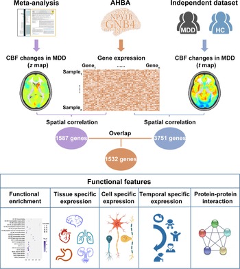
To elucidate such mechanisms, we initially determined CBF changes in MDD by performing a comprehensive neuroimaging meta-analysis as well as examining group CBF differences in an independent large sample of 133 MDD patients and 133 healthy controls (HC). Notably, compared with the Wang et al., meta-analysis including only ASL studies (Wang & Yang, Reference Wang and Yang2022), our meta-analysis included studies from both ASL and PET/SPECT. In combination with the AHBA, transcriptome-neuroimaging spatial correlation analyses were then conducted to identify genes whose expression patterns were associated with CBF changes in MDD. Finally, a set of post hoc analyses [i.e. functional enrichment, specific expression, behavioral relevance, and protein-protein interaction (PPI) analyses] were carried out for the identified genes to describe their functional features. A schematic overview of the study design and analysis pipeline is shown in Fig. 1.
A schematic overview of the study design and analysis pipeline. AHBA, Allen Human Brain Atlas; CBF, cerebral blood flow; HC, healthy controls; MDD, major depressive disorder.

Materials and methods
Literature search and selection
Our meta-analysis was conducted according to the Preferred Reporting Items for Systematic Reviews and Meta-Analyses (PRISMA) guidelines (Moher, Liberati, Tetzlaff, Altman, & Group, Reference Moher, Liberati, Tetzlaff and Altman2009). A systematic search was performed independently by two investigators (X.S. and W.H.) to determine relevant studies published in PubMed and Web of Science before 19 February 2021. This meta-analysis protocol was pre-registered in PROSPERO (https://www.crd.york.ac.uk/PROSPERO/, registration number: CRD42021250728). The search terms along with the inclusion and exclusion criteria can be found in the Supplementary materials. The detailed study selection process is shown in online Supplementary Fig. S1. For each included study, we recorded the following information: scanner, sample size, sex, age, symptom severity, diagnostic criteria, and illness duration.
Neuroimaging meta-analysis of group CBF differences
Voxel-wise meta-analysis of CBF differences between MDD patients and HC was conducted using Seed-based d Mapping with Permutation of Subject Images (SDM-PSI, version 6.21) (https://www.sdmproject.com). In contrast to the traditional activation likelihood estimation (ALE) method, SDM-PSI enables investigators to combine both peak coordinates and statistical parametric maps and uses standard effect size and variance-based meta-analytic calculations. The SDM method has been described in detail elsewhere (Albajes-Eizagirre et al., Reference Albajes-Eizagirre, Solanes, Fullana, Ioannidis, Fusar-Poli, Torrent and Radua2019a). First, we extracted peak coordinates and corresponding effect sizes (e.g. t values) of clusters with significant CBF differences between MDD patients and HC from each study. Coordinates reported in the Talairach space were converted to the Montreal Neurological Institute (MNI) space using the matrix transformations proposed by Lancaster (Lancaster et al., Reference Lancaster, Tordesillas-Gutierrez, Martinez, Salinas, Evans, Zilles and Fox2007); z or p values were converted to t values using SDM online conversion utilities (https://www.sdmproject.com/utilities/?show=Statistics). Then, a standard MNI map of CBF differences between MDD patients and HC was separately created for each study using a Gaussian kernel of 20 mm full width at half maximum (Radua et al., Reference Radua, Mataix-Cols, Phillips, El-Hage, Kronhaus, Cardoner and Surguladze2012). Next, these maps were combined using a standard random-effects model accounting for sample size, intra-study variability and between-study heterogeneity, resulting in a final map of group CBF differences (z map) for all included studies. Considering the relatively small number of included studies, we reported results using a voxel-wise threshold of p < 0.005 combined with a cluster size threshold of 10 voxels to optimally balance Types I and II error rates (Lieberman & Cunningham, Reference Lieberman and Cunningham2009; Radua et al., Reference Radua, Mataix-Cols, Phillips, El-Hage, Kronhaus, Cardoner and Surguladze2012). Several supplementary analyses were pursued to test the robustness and reliability of our meta-analysis results. Detailed steps are described in the Supplementary materials.
Participants in the independent dataset
Group CBF differences were also tested in an independent sample of 133 MDD patients and 133 well-matched HC. MDD patients were enrolled consecutively from the inpatient and outpatient departments of Hefei Fourth People's Hospital. HC were recruited from the local community via poster advertisements. This study was conducted in accordance with the Declaration of Helsinki and was approved by the ethics committee of The First Affiliated Hospital of Anhui Medical University (5101152). Written informed consent was obtained from all participants after they had been given a complete description of the study. Inclusion and exclusion criteria for all participants are described in the Supplementary materials.
MRI data acquisition and CBF analysis
MRI data were acquired on a 3.0-Tesla MR system (Discovery MR750w, General Electric, Milwaukee, WI, USA). The resting-state perfusion imaging was performed using a pseudo-continuous ASL sequence with a 3D fast spin-echo acquisition and background suppression. Detailed scan sequences and parameters can be found in the Supplementary materials. Three ASL difference images were calculated by subtracting the label images from the control images and then averaged. Next, CBF was quantified by applying a single-compartment model (Buxton et al., Reference Buxton, Frank, Wong, Siewert, Warach and Edelman1998) to the mean ASL difference and proton-density-weighted reference images (Xu et al., Reference Xu, Rowley, Wu, Alsop, Shankaranarayanan, Dowling and Johnson2010; Zhu et al., Reference Zhu, Zhuo, Qin, Xu, Xu, Liu and Yu2015, Reference Zhu, Zhuo, Xu, Liu, Qin and Yu2017; Zhuo et al., Reference Zhuo, Zhu, Qin, Qu, Ma and Yu2017). The Statistical Parametric Mapping software (SPM12, http://www.fil.ion.ucl.ac.uk/spm) was used to normalize the CBF images into the MNI space. The detailed steps are described in the Supplementary materials. For the independent dataset, CBF differences between MDD patients and HC were examined using a two-sample t test in a voxel-wise manner, resulting in a statistical t map. Multiple comparisons were corrected using the cluster-level family-wise error (FWE) method, resulting in a cluster defining threshold of p < 0.001 and a corrected cluster significance of p < 0.05.
Brain gene expression data processing
Brain gene expression data were obtained from the downloadable AHBA dataset (http://www.brain-map.org) (Hawrylycz et al., Reference Hawrylycz, Lein, Guillozet-Bongaarts, Shen, Ng, Miller and Jones2012, Reference Hawrylycz, Miller, Menon, Feng, Dolbeare, Guillozet-Bongaarts and Lein2015). The dataset was derived from six human post-mortem donors (online Supplementary Table S1). The original expression data of more than 20 000 genes at 3702 spatially distinct brain tissue samples were processed using a newly proposed pipeline (Arnatkeviciute, Fulcher, & Fornito, Reference Arnatkeviciute, Fulcher and Fornito2019). The detailed steps can be found in the Supplementary materials. After these processing procedures, we obtained normalized expression data of 5013 genes for 1280 tissue samples. Because our neuroimaging meta-analysis was performed within a gray matter mask provided by the SDM-PSI, we further restricted our analyses to the samples within this mask, resulting in a final sample × gene matrix of 894 × 5013.
Transcription-neuroimaging association analysis
To derive the group CBF difference in a given brain tissue sample, we drew a spherical region (radius = 3 mm) centered at the MNI coordinate of this sample and extracted the average z value of voxels within the sphere from the meta-analysis z map. Then, cross-sample (894 samples) Pearson's correlations between gene expression and z values were performed in a gene-wise manner (5013 genes), yielding 5013 correlation coefficients. Multiple comparisons were corrected using the Bonferroni method (p < 0.05/5013 = 9.974 × 10⁻⁶). Likewise, the above-described analysis procedure was conducted for the t map in the independent dataset. Then, only genes with significant spatial correlations with both the meta-analysis z map and the independent dataset t map were considered as the genes whose expression levels were associated with CBF changes in MDD patients. To further test whether the number of the identified genes was significantly greater than the random level, a spatially-constrained permutation test was conducted to establish the significance of our results. The detailed steps are described in the Supplementary materials.
Gene enrichment analysis
A series of enrichment analyses were conducted for the identified genes associated with CBF changes in MDD patients. First, functional annotation was carried out with use of the ToppGene portal (https://toppgene.cchmc.org/) (Chen, Bardes, Aronow, & Jegga, Reference Chen, Bardes, Aronow and Jegga2009). Gene ontology (GO) was used to determine the biological functions including molecular functions (MFs), biological processes (BPs), and cellular components (CCs). The disease database was adopted to determine the related diseases. Second, we used online tissue-specific expression analysis (http://genetics.wustl.edu/jdlab/tsea/) and cell type-specific expression analysis (http://genetics.wustl.edu/jdlab/csea-tool-2/) (Dougherty, Schmidt, Nakajima, & Heintz, Reference Dougherty, Schmidt, Nakajima and Heintz2010) tools to conduct tissue, cell type, and temporal specific expression analyses, with the aim of determining the specific tissues, cortical cell types, and developmental stages in which these genes were overrepresented. A specificity index probability (pSI) was used to determine how likely a gene was to be specifically expressed (Xu, Wells, O'Brien, Nehorai, & Dougherty, Reference Xu, Wells, O'Brien, Nehorai and Dougherty2014) and four pSI thresholds (0.05, 0.01, 0.001, and 0.0001) were employed in this analysis. Finally, we examined the overlap between the genes associated with CBF changes in MDD patients found in the current study and MDD-associated genes in the MalaCards database (https://www.malacards.org/) (Rappaport et al., Reference Rappaport, Twik, Plaschkes, Nudel, Iny Stein, Levitt and Lancet2017), using 20 737 genes with unique Entrez IDs in the AHBA as the background list. For the aforementioned enrichment analyses, Fisher's exact tests were used to test their significance. Multiple testing was corrected using the Benjamini and Hochberg method for false discovery rate (FDR-BH correction) with a corrected p value (q) of 0.05.
Behavioral relevance analysis
To capture the behavioral relevance of the genes related to CBF changes in MDD patients, we tested their associations with behavioral domains from the Neurosynth (https://neurosynth.org/), a well-validated and publicly available platform for meta-analysis of neuroimaging literature (Yarkoni, Poldrack, Nichols, Van Essen, & Wager, Reference Yarkoni, Poldrack, Nichols, Van Essen and Wager2011). Detailed steps are provided in the Supplementary materials.
Protein-protein interaction analysis
PPI analysis was performed with STRING v11.0 (https://string-db.org/) to determine whether the genes associated with CBF changes in MDD patients could construct a PPI network with a highest confidence interaction score of 0.9. Genes with the top 10% highest degree values (i.e. the number of edges connected to a gene) were defined as hub genes. In addition, the Human Brain Transcriptome database (http://hbatlas.org/) was employed to delineate the spatial-temporal expression trajectory of hub genes with the highest degree values.
Results
Meta-analysis of CBF changes in MDD patients
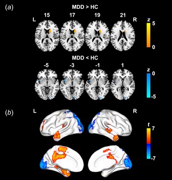
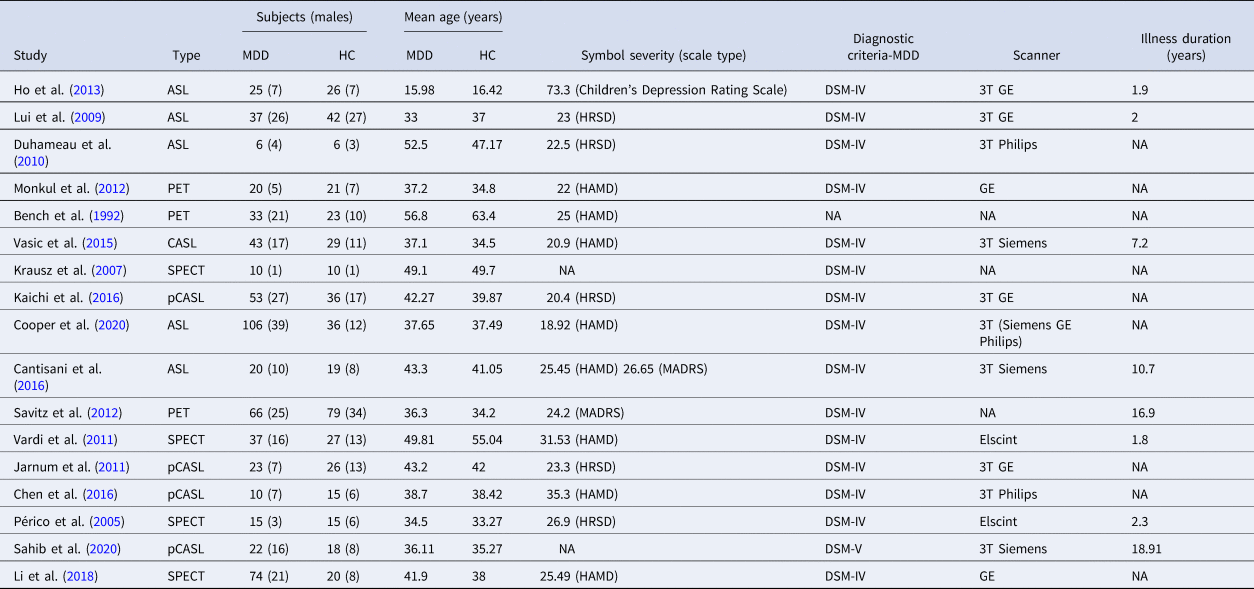
After a comprehensive literature search and selection, our neuroimaging meta-analysis included 600 MDD patients and 448 HC from 17 studies. The demographic and clinical characteristics of these participants are shown in Table 1. Compared with HC, MDD patients exhibited increased CBF in the right caudate and decreased CBF in the left insula and right calcarine sulcus (Fig. 2a and online Supplementary Table S2). Moreover, jackknife sensitivity analysis (82–88% consistency) (online Supplementary Table S3), publication bias analysis including small-study effect and excess significance tests (p > 0.05) (online Supplementary Table S4), and heterogeneity analysis (I 2 < 50%) (online Supplementary Table S5) demonstrated the robustness and reliability of our meta-analysis results.
(a) Brain regions with CBF differences between MDD patients and HC identified by the neuroimaging meta-analysis. (b) Brain regions with CBF differences between MDD patients and HC in the independent dataset. Warm and cold colors denote increased and decreased CBF in MDD patients, respectively. CBF changes in the subcortical regions are not shown. CBF, cerebral blood flow; HC, healthy controls; MDD, major depressive disorder; L, left; R, right.

Demographic and clinical characteristics of the 17 studies in the meta-analysis

ASL, arterial spin labeling; CASL, continuous arterial spin labeling; DSM, diagnostic statistical manual of mental disorders; GE, General Electric Company; HAMD, Hamilton Rating Scale for Depression; HC, healthy controls; HRSD, Hamilton Rating Scale for Depression; MADRS, Montgomery-Asberg Depression Rating Scale; MDD, major depressive disorder; NA, not available; pCASL, pseudo-continuous arterial spin labeling; PET, positron emission tomography; SPECT, single photon emission computed tomography
CBF changes in MDD patients in the independent dataset
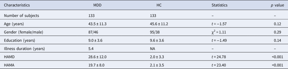
The demographic and clinical characteristics of the independent sample are presented in Table 2. There were no significant differences in age, gender, and education between MDD patients and HC. The voxel-wise two-sample t test revealed a mix of increased and decreased CBF in MDD patients (Fig. 2b and online Supplementary Table S6). Specifically, MDD patients showed increased CBF in the bilateral middle cingulate gyrus/bilateral supplementary motor area/left posterior cingulate gyrus/left precuneus, left putamen, right temporal pole, left middle temporal gyrus, right superior temporal gyrus, bilateral thalamus, and left middle frontal gyrus relative to HC (p < 0.05, cluster-level FWE corrected). In addition, MDD patients showed decreased CBF in the bilateral occipital cortex extending to bilateral parietal cortex in comparison with HC (p < 0.05, cluster-level FWE corrected).
Demographic and clinical characteristics of the independent sample

HAMA, Hamilton Rating Scale for Anxiety; HAMD, Hamilton Rating Scale for Depression; HC, healthy controls; MDD, major depressive disorder; NA, not available
Genes associated with CBF changes in MDD patients
Leveraging transcriptome-neuroimaging spatial correlation analyses, we found that the meta-analysis z map and the independent dataset t map were respectively associated with expression measures of 1587 and 3751 genes (p < 0.05, Bonferroni corrected), with 1532 overlap genes (Supplementary file 1). These overlap genes, whose expression levels were considered to associate with CBF changes in MDD patients, were used for further analyses. The spatially-constrained permutation test showed that none out of 5000 permutations resulted in more genes than those identified using the real data (P perm < 0.0002), indicating that our results were different from random. Moreover, we observed significant overlaps between the genes in the main analysis and those identified using two other differential stability (DS) thresholds of 40% (overlap ratio: 97.80%) and 60% (overlap ratio: 96.42%) (online Supplementary Table S7 and file 2).
Gene functional enrichment
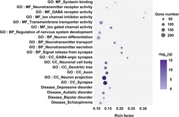
To characterize the biological functions and diseases of the genes associated with CBF changes in MDD patients, we performed functional enrichment analyses using the ToppGene portal. The results of functional enrichment are listed in Supplementary file 3 and are depicted in Fig. 3. With regard to GO, these genes were enriched for MFs including syntaxin binding, neurotransmitter receptor activity, GABA receptor activity, ion channel inhibitor activity, transmembrane transporter activity, and ion gated channel activity; for BPs including regulation of nervous system development, neuron differentiation, neurotransmitter transport, neurotransmitter secretion, and signal release from synapse; and for CCs including neuron projection, neuronal cell body, axon, dendritic tree, synapse, and GABA-ergic synapse. As to diseases, these genes were found to be enriched for several common mental disorders including depressive disorder, autistic disorder, bipolar disorder, and schizophrenia.
Functional enrichment of the genes associated with CBF changes in MDD patients. For the bubble chart, the x-axis represents the rich factor and the y-axis represents items from the GO and disease databases. The rich factor refers to the ratio of the number of the significant genes annotated to the item to the number of all genes annotated to the item. The bubble size denotes the number of genes overlapping with those belonging to each item, and the bubble color represents the −log10(q). BP, biological process; CBF, cerebral blood flow; CC, cellular component; GO, gene ontology; MDD, major depressive disorder; MF, molecular function.

Tissue, cell type, and temporal specific expression
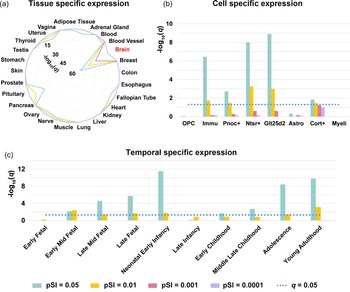
The specific expression results of the 1532 genes related to CBF changes in MDD patients are listed in Supplementary file 4 and are illustrated in Fig. 4. Tissue specific expression analysis demonstrated that these genes were specifically expressed in the brain tissue (Fig. 4a). Cell type specific expression analysis revealed that these genes were specifically expressed in immune cells and multiple types of neurons including Pnoc+, Ntsr+, Glt25d2, and Cort+ (Fig. 4b). Temporal specific expression analysis showed that these genes were preferentially expressed during early mid-fetal, late mid-fetal, late fetal, neonatal and early infancy, early childhood, middle and late childhood, adolescence, and young adulthood (Fig. 4c).
Specific expression of the genes associated with CBF changes in MDD patients. (a) Tissue specific expression. (b) Cell specific expression. (c) Temporal specific expression. Astro, astrocytes; CBF, cerebral blood flow; Cort+, corticosterone-expressing neurons; Glt25d2, corticopontine neurons; Immu, immune cells; MDD, major depressive disorder; Myeli, myelinating oligodendrocytes; Ntsr+, corticothalamic neurons; OPC, oligodendrocyte progenitor cells; Pnoc+, prepronociceptin-expressing neurons.

Overlap with MDD-associated genes
Fisher's exact test revealed that the 1532 genes associated with CBF changes in MDD patients found in the current study significantly overlapped with the 110 MDD-associated genes in the Malacards database (17 overlap genes, odds ratio = 2.10, p = 6.26 × 10⁻³).
Behavioral relevance
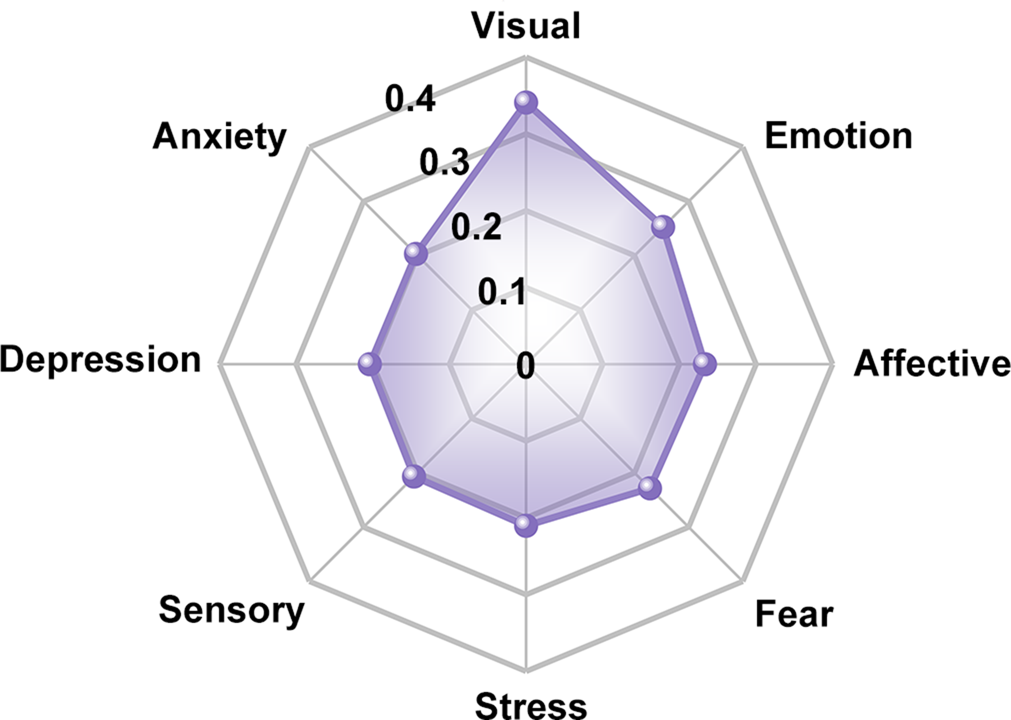
By linking gene expression with behavioral domains via the Neurosynth, we found that the genes associated with CBF changes in MDD patients were correlated with multiple behavioral terms including visual, emotion, affective, fear, stress, sensory, depression, and anxiety (Fig. 5).
Behavioral relevance of the genes associated with CBF changes in MDD patients. The radial array numbers represent average absolute correlation coefficients between expression measures of the identified genes and activation values of behavioral terms from the Neurosynth. CBF, cerebral blood flow; MDD, major depressive disorder.

PPI network and hub genes
PPI analysis revealed that 604 genes from the 1532 genes could construct an interconnected PPI network (online Supplementary Fig. S3A). This network consisted of 3582 edges, which was significantly higher than expected (p = 9.6 × 10⁻¹²). 60 genes with the top 10% highest degree values were defined as hub genes (Supplementary file 5). In addition, we delineated the spatial-temporal expression trajectory of three hub genes with the highest degree values (i.e. GNG2, GNB4, and GNG4) (online Supplementary Fig. S3B).
Discussion
To our knowledge, this is the first study to investigate the genetic mechanisms of CBF changes in MDD using a combined analysis of brain imaging and gene expression data. Neuroimaging meta-analysis of previous literature and group comparison analysis in an independent dataset consistently demonstrated that MDD patients had increased CBF in the reward circuitry and default-mode network and decreased CBF in the visual system. Moreover, transcriptome-neuroimaging correlation analysis revealed that these CBF changes were spatially associated with expression of 1532 genes, which were enriched for important MFs, BPs, and CCs of the cerebral cortex as well as several common mental disorders. Concurrently, these genes were specifically expressed in the brain tissue, in immune cells and neurons, and during nearly all developmental stages. Regarding behavioral relevance, these genes were associated with domains involving emotion and sensation. In addition, these genes could construct a PPI network supported by 60 putative hub genes with functional significance. Altogether, our findings suggest that CBF changes in MDD may be a consequence of complex interactions of a wide range of genes with diverse functional features, confirming the polygenic nature of this illness.
The neuroimaging meta-analysis revealed that MDD patients presented with increased CBF in the caudate and decreased CBF in the insula and calcarine sulcus relative to HC. In the independent dataset, MDD patients showed increased CBF in the middle cingulate gyrus, supplementary motor area, posterior cingulate gyrus/precuneus, putamen, temporal cortex, thalamus and middle frontal gyrus, as well as decreased CBF predominantly in the occipital cortex. These results can be summarized as hyper-perfusion in the reward circuitry [the caudate, putamen, and middle frontal gyrus (Clery-Melin, Jollant, & Gorwood, Reference Clery-Melin, Jollant and Gorwood2019)] and default-mode network (the posterior cingulate gyrus/precuneus, lateral temporal cortex, and thalamus [Buckner & DiNicola, Reference Buckner and DiNicola2019; Raichle, Reference Raichle2015)] and hypo-perfusion in the visual system (the occipital cortex), indicating a perfusion redistribution in the brain of MDD patients. It is well established that MDD is a clinically heterogeneous disorder characterized by a mixture of emotional, cognitive, and neurovegetative symptoms (Malhi & Mann, Reference Malhi and Mann2018). Anhedonia represents one of the fundamental emotional symptoms in patients suffering from MDD, which is thought to associate with impaired reward processing possibly arising from dysfunction in the reward circuitry (Hoflich, Michenthaler, Kasper, & Lanzenberger, Reference Hoflich, Michenthaler, Kasper and Lanzenberger2019; Li et al., Reference Li, Yang, Zhu, Zhou, Han, Yin and Chen2018; Nestler & Carlezon, Reference Nestler and Carlezon2006; Pizzagalli et al., Reference Pizzagalli, Holmes, Dillon, Goetz, Birk, Bogdan and Fava2009; Russo & Nestler, Reference Russo and Nestler2013). For example, Pizzagalli et al., found caudate dysfunction during reward processing as well as a link between caudate volume and anhedonia in MDD patients (Pizzagalli et al., Reference Pizzagalli, Holmes, Dillon, Goetz, Birk, Bogdan and Fava2009). Tao and colleagues reported that MDD patients exhibited abnormal function in the ‘hate circuit’ involving the putamen and prefrontal cortex, which may reflect reduced cognitive control over negative feelings toward both self and others (Tao et al., Reference Tao, Guo, Ge, Kendrick, Xue, Liu and Feng2013). Rumination, defined as repetitive thinking about negative information, is a characteristic of MDD (Whitmer & Gotlib, Reference Whitmer and Gotlib2013). The default-mode network is classically considered an ‘intrinsic’ system, specializing in internally oriented cognitive processes such as daydreaming, reminiscing and future planning (Yeshurun, Nguyen, & Hasson, Reference Yeshurun, Nguyen and Hasson2021). There is empirical evidence from functional neuroimaging pointing to an intimate link between rumination and activations of the default-mode network (Zhou et al., Reference Zhou, Chen, Shen, Li, Chen, Zhu and Yan2020). Our observation of decreased CBF in the visual system is coherent with previous reports (Bonte et al., Reference Bonte, Trivedi, Devous, Harris, Payne, Weinberg and Haley2001; Nagafusa et al., Reference Nagafusa, Okamoto, Sakamoto, Yamashita, Kawaguchi, Higuchi and Matsuda2012). It is largely known that the visual system is not only implicated in visual processing (Prasad & Galetta, Reference Prasad and Galetta2011), but also engaged by higher level cognitive processes (Roelfsema & de Lange, Reference Roelfsema and de Lange2016). Earlier research indicates that functional deficit in the visual system may be an initiating factor for cognitive impairment in MDD patients (Li et al., Reference Li, Xu, Cao, Gao, Wang, Wang and Zhang2013). In addition, we observed decreased CBF in the insula, which plays a pivotal role in interoceptive awareness, emotional responses, and empathic processes (Menon & Uddin, Reference Menon and Uddin2010). This finding complements and extends prior literature on structural and functional impairments in this region in MDD (Liu et al., Reference Liu, Xu, Xu, Wang, Zhao, Lv and Du2010; Stratmann et al., Reference Stratmann, Konrad, Kugel, Krug, Schoning, Ohrmann and Dannlowski2014). Collectively, our data suggest that there exists a cerebral perfusion redistribution across the reward circuitry, default-mode network, and visual system that might account for the clinical heterogeneity of MDD.
More importantly, leveraging transcription-neuroimaging spatial association analysis, we identified 1532 genes whose expression patterns were correlated with CBF changes in MDD. Functional enrichment analyses demonstrated that these genes were mainly enriched for ion channel (ion channel inhibitor activity and ion gated channel activity), synapse (syntaxin binding, signal release from synapse, and GABA-ergic synapse), neuron (neuron differentiation, neuronal cell body, and neuron projection), and neurotransmitter system (neurotransmitter receptor activity, GABA receptor activity, neurotransmitter transport, and neurotransmitter secretion). Ion channels are important for generating membrane potential, neuronal growth and differentiation, signal transduction, and neurotransmitter release (Kumar, Kumar, Jha, Jha, & Ambasta, Reference Kumar, Kumar, Jha, Jha and Ambasta2016; Moody & Bosma, Reference Moody and Bosma2005; Smith & Walsh, Reference Smith and Walsh2020). Previous studies have suggested that ion channels, such as low-voltage-sensitive T-type calcium channel and potassium channel Kir4.1, hold promise as therapeutic targets for depression (Hashimoto, Reference Hashimoto2019). It is generally accepted that brain function relies on the ability of neurons to communicate with each other; inter-neuronal communication primarily takes place at synapses, where information from one neuron is rapidly conveyed to a second neuron (Pereda, Reference Pereda2014). Basic studies have documented that ketamine, a rapid antidepressant, can induce synaptogenesis and reverse synaptic deficits caused by chronic stress, highlighting the role of synaptic dysfunction in depression and its potential as a therapeutic target (Duman & Aghajanian, Reference Duman and Aghajanian2012). It is quite apparent that neuronal impairment and maldevelopment may be central to the neurology of depression (Krishnan & Nestler, Reference Krishnan and Nestler2008; Willner, Scheel-Krüger, & Belzung, Reference Willner, Scheel-Krüger and Belzung2013). Besides, neurons are a key component of the neurovascular unit, which serves as the structural basis of the coupling between CBF and neuronal activity. Central to brain health, neurovascular interactions have emerged as essential contributors to brain development and are vital for maintaining the homeostasis of the brain internal milieu (Iadecola, Reference Iadecola2017). Accumulating data have indicated the occurrence of neurovascular coupling disruption in major psychiatric disorders including MDD (Segarra, Aburto, Hefendehl, & Acker-Palmer, Reference Segarra, Aburto, Hefendehl and Acker-Palmer2019). Extensive research indicates that dysfunction of neurotransmitter systems (e.g. GABA, glutamate, and dopamine) has been implicated in the pathophysiology of MDD (Choudary et al., Reference Choudary, Molnar, Evans, Tomita, Li, Vawter and Jones2005; Grace, Reference Grace2016; Klempan et al., Reference Klempan, Sequeira, Canetti, Lalovic, Ernst, ffrench-Mullen and Turecki2009). Moreover, SAGE-217, a positive allosteric modulator of GABA type A receptors, has been proven to be effective for the treatment of MDD (Gunduz-Bruce et al., Reference Gunduz-Bruce, Silber, Kaul, Rothschild, Riesenberg, Sankoh and Kanes2019). With respect to diseases, the genes related to CBF changes in MDD were found to be enriched for several mental disorders including depressive disorder, autistic disorder, bipolar disorder and schizophrenia, indicative of some common genetic mechanisms contributing to these illnesses.
Specific expression analyses showed that the genes associated with CBF changes in MDD were specifically expressed in the brain tissue, in immune cells and neurons, and during nearly all developmental stages. It is often assumed that immune cells (e.g. microglia) have the potential to regulate the development, structure, and function of neuronal networks in the brain (Garaschuk & Verkhratsky, Reference Garaschuk and Verkhratsky2019). In addition, immune cells have been evidenced to involve the establishment of the MDD model, emphasizing the neuroimmune mechanisms of depression (Hodes, Kana, Menard, Merad, & Russo, Reference Hodes, Kana, Menard, Merad and Russo2015). The specific expression in neurons echoes the aforementioned functional enrichment results. The temporal specific expression indicates that these genes exert an enduring effect on MDD-related CBF changes throughout nearly all cortical developmental stages. Regarding behavioral relevance, the genes related to CBF changes in MDD were associated with domains involving emotion and sensation. Deficits in these behavioral domains have been widely reported in MDD (Fam, Rush, Haaland, Barbier, & Luu, Reference Fam, Rush, Haaland, Barbier and Luu2013; Rottenberg, Reference Rottenberg2017; Salmela et al., Reference Salmela, Socada, Soderholm, Heikkila, Lahti, Ekelund and Isometsa2021). Of note, these genes could construct a PPI network supported by 60 putative hub genes with functional significance for understanding the pathology and treatment of MDD. For example, several G protein related genes (i.e. GNG2, GNB4, and GNG4) encode G protein coupled receptors (GPCRs), which could mediate the eukaryotic repertoire for cell communication and signal transduction. GPCRs are considered to be implicated in MDD (Mantas, Saarinen, Xu, & Svenningsson, Reference Mantas, Saarinen, Xu and Svenningsson2022) and several novel antidepressants targeting GPCRs have been developed (Borroto-Escuela et al., Reference Borroto-Escuela, Ambrogini, Narvaez, Di Liberto, Beggiato, Ferraro and Fuxe2021; Mantas et al., Reference Mantas, Saarinen, Xu and Svenningsson2022).
Several limitations should be taken into account when interpreting our findings. First, most of the MDD patients in both the neuroimaging meta-analysis and the independent dataset were antidepressant-medicated and chronic, which may introduce confounds of antidepressant medication and illness duration. Future studies with drug-naïve first-episode patients with MDD are needed to eliminate these confounds and verify the preliminary findings. Second, the meta-analysis z map cannot reflect the nature and extent of whole-brain CBF differences between MDD patients and HC, because only peak coordinates and corresponding effect sizes of significant clusters reported in the previous literature were used. Third, the gene expression data were obtained from post-mortem brains, while the neuroimaging data were collected from living brains. Given the mounting evidence of conservative gene expression across individuals (Hawrylycz et al., Reference Hawrylycz, Miller, Menon, Feng, Dolbeare, Guillozet-Bongaarts and Lein2015; Zeng et al., Reference Zeng, Shen, Hohmann, Oh, Bernard, Royall and Jones2012), we set a DS threshold to focus on the genes with more conserved expression patterns, such that our transcription-neuroimaging spatial association analysis is feasible. However, some genes with great inter-subject variability in expression profiles would be missed. Fourth, considering limited gene expression data in the right hemisphere and substantial gene expression divergence between cortical and subcortical regions, we only included the tissue samples in the left cerebral cortex, which may have introduced potential biases. Finally, our neuroimaging meta-analysis results cannot survive multiple comparison correction. Instead, we reported our results by using a voxel-wise threshold of p < 0.005 combined with a cluster size threshold of 10 voxels to optimally balance Types I and II error rates following prior recommendation (Radua et al., Reference Radua, Mataix-Cols, Phillips, El-Hage, Kronhaus, Cardoner and Surguladze2012).
In conclusion, our data showed a cerebral perfusion redistribution in MDD, characterized by increased CBF in the reward circuitry and default-mode network and decreased CBF in the visual system. Further transcriptome-neuroimaging correlation analyses demonstrated that these CBF changes were spatially associated with expression of 1532 genes with diverse functional features. Our findings may not only offer unique insight into the genetic mechanisms of CBF changes in MDD, but also inform novel treatment approaches targeting the molecular substrates underlying brain phenotypes of this disorder.
Supplementary material
The supplementary material for this article can be found at https://doi.org/10.1017/S0033291722003750.
Acknowledgements
This work was supported by the National Natural Science Foundation of China (grant number: 82071905), the Natural Science Foundation of Anhui Province (grant number: 2208085MH257), the Outstanding Youth Support Project of Anhui Province Universities (grant number: gxyqZD2022026) and the Scientific Research Foundation of Anhui Medical University (grant number: 2022xkj143). We thank the Allen Institute for Brain Science founders and staff who supplied the brain expression data.
Conflict of interest
The authors declare no conflict of interests.