Introduction
The jugular bulb is the junction of the sigmoid sinus and the internal jugular vein. Inter-individual variation in its size and location are thought to be caused by venous events during the first few decades of life.Reference Friedmann, Eubig, Winata, Pramanik, Merchant and Lalwani1–Reference Friedmann, Eubig, McGill, Babb, Pramanik and Lalwani3 Abnormalities of the jugular bulb, including high jugular bulb, jugular bulb diverticulum and jugular bulb dehiscence, are widely recognised. However, high jugular bulb is considered to be the most common and clinically significant abnormality.Reference Fujimoto, Ito, Ishimoto and Iwasaki4–Reference Hourani, Carey and Yousem7 Temporal bone and radiological studies suggest that the incidence of high jugular bulb ranges from 3 to 24 per cent, although clinical symptoms may not be present.Reference Vachata, Petrovicky and Sames5, Reference Wadin, Thomander and Wilbrand8–Reference Kuhn, Friedmann, Winata, Eubig, Pramanik and Kveton10
Jugular bulb abnormalities have been linked to a variety of cochleovestibular symptoms, depending on their impact on surrounding structures.Reference Friedmann, Eubig, Winata, Pramanik, Merchant and Lalwani2 The jugular bulb may erode into the middle ear or the vestibular or cochlear aqueduct, or may affect the endolymphatic sac or duct.Reference Friedmann, Eubig, Winata, Pramanik, Merchant and Lalwani1, Reference Vachata, Petrovicky and Sames5 Dehiscence of a high jugular bulb and the vestibular aqueduct, as well as jugular bulb interference with the endolymphatic sac, has been implicated in producing Ménière's disease-like symptoms, such as hearing loss, vertigo and tinnitus.Reference Bilgen, Kirazli, Ogut and Totan6, Reference Hourani, Carey and Yousem7, Reference Couloigner, Grayeli, Bouccara, Julien and Sterkers11–Reference Manzari14
Research has focused on how the vestibular aqueduct is affected by jugular bulb abnormalities. However, Wadin et al. reported that the cochlear aqueduct was the structure primarily affected by a high jugular fossa.Reference Wadin, Thomander and Wilbrand8 Furthermore, other studies have demonstrated that impingement on the vestibular aqueduct does not cause endolymphatic hydrops, which is typically present in Ménière's disease patients. Because the cochlear aqueduct is known to play a significant role in the balance between perilymphatic, endolymphatic and cerebrospinal fluid,Reference Proctor, Proctor and Proctor15 and changes in pressure gradients of these fluids have been linked to endolymphatic hydrops,Reference Bohmer and Dillier16 we hypothesised that interference of a high jugular bulb with the cochlear aqueduct may play a role in development of endolymphatic hydrops and Ménière's disease.
Materials and methods
Institutional Review Board approval was obtained for this retrospective review of patients treated for definite Ménière's disease in 2011. Inclusion criteria were age 18 years or older, no other potential cause of dizziness or hearing loss (such as previous trauma or surgery) and classification as definite Ménière's patients.17 Included patients were refractory to at least a three month trial of lifestyle modifications with diuretic therapy (if no contraindications existed). Non-surgical treatment options were discussed with all patients. Patients elected to undergo either endolymphatic sac and posterior fossa decompression or surgical labyrinthectomy. Only those with a pre-operative computed tomography (CT) scan performed at St. Luke's Hospital (Maumee, Ohio) through the Ohio Hearing and Balance Institute were included because the use of a single picture archiving and communication system minimised variations in CT quality and radiographic measurements. A total of 29 patients were eligible for inclusion, but 3 of these were excluded because of poor scan quality or because their data could not be accessed. Therefore, 26 patients were included in the final analysis. A GE Light Speed VCT 64-detector CT (GE Amersham Healthcare System, Milwaukee, Wisconsin, USA) was used to scan the temporal bone in this patient group prior to elective surgical intervention. Demographic information and complete audiometric data for both affected and contralateral ears were collected.
Radiographs were reviewed to collect anatomical information, including the height and position of the jugular bulb (medial or intermediate, based on its position relative to the basal turn of the cochlea or the round window). A high jugular bulb was defined as reaching or rising above the basal turn of the cochlea, since the cochlear basal turn is easily identified in most cases.Reference Wadin, Thomander and Wilbrand8, Reference Friedmann, Le, Pramanik and Lalwani18, Reference Low, Fenton, Fagan and Gibson19 The largest dimensions of the jugular bulb (anterior to posterior and medial to lateral) were recorded for both affected and contralateral ears. The area of the jugular bulb was calculated as described previously by Friedmann et al. Reference Friedmann, Eubig, Winata, Pramanik, Merchant and Lalwani1, Reference Friedmann, Eubig, McGill, Babb, Pramanik and Lalwani3 Additionally, visible encroachment of the cochlear or vestibular aqueduct were noted. Unusual anatomical findings apparent on radiographs (bony dehiscence or presence of a diverticulum) were also recorded.
Statistical analysis
Descriptive statistics were used to report demographic data. The height and width of the jugular bulb in affected and contralateral ears were compared using Student's t-tests. The location of the jugular bulb in relation to the midpoint of the lumen at the inferior limit of the cochlea in the affected ear (left vs right) and unaffected ear (affected vs contralateral) were compared using Fisher's exact test. Encroachment of the cochlear and vestibular aqueduct was analysed according to the affected ear (left vs right) and the position of the jugular bulb (high vs normal) using Fisher's exact test. Unusual findings in the affected ears were compared for sidedness (left vs right) and for location of the jugular bulb (high vs normal) using Fisher's exact test. Patients affected bilaterally were excluded from all analyses comparing contralateral and affected ears, but included in all analyses comparing right and left ears. All analyses were performed using R version 2.12.2 (Vienna, Austria; http://www.r-project.org).20 Tests were declared statistically significant at a p value of < 0.05.
Results
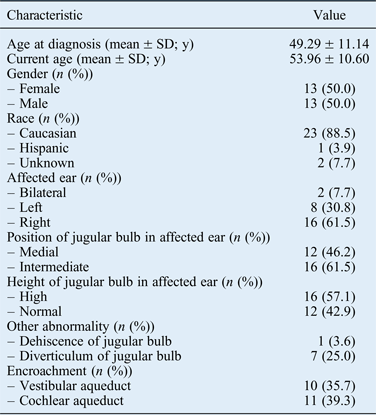
A total of 26 patient charts were reviewed in this study, and 50 per cent of patients were female. The average age at diagnosis was 49.29 ± 11.14 years, and the average age at the time of the study was 53.96 ± 10.60 years. Only two patients were affected bilaterally, and 61.5 per cent of patients experienced symptoms in the right ear only. The position of the jugular bulb in the affected ear was judged to be medial in 46.2 per cent of patients, whereas 61.5 per cent of radiographs indicated an intermediate position (Table I). Importantly, the jugular bulb was judged to be high in 57.1 per cent of affected ears.
Patient demographics

y = years
Audiograms of all affected and unaffected ears were compared to determine whether jugular bulb abnormalities affect hearing loss and, if so, whether the effect is related to sidedness. First, mean hearing loss (determined by audiogram; in decibels) in the contralateral, or unaffected, ears was compared between patient groups affected in the right or left ears. No significant difference in mean hearing loss was detected at any frequency (data not shown).
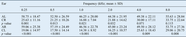
Similarly, hearing loss was compared between patients affected on the right side and those affected on the left. No significant differences in hearing were found between patients with affected right and affected left ears at any frequency. The mean hearing loss in affected ears was then compared with the mean hearing loss recorded for the contralateral ears. Patients whose left ears were affected showed significant differences in mean hearing between the affected and non-affected sides at 0.25, 0.5, 1.0 and 2.0 kHz. Patients affected on the right side showed significant differences in mean hearing at all frequencies tested compared with their contralateral ears (Table II).
Hearing loss in affected and contralateral ears
AL = affected left; CR = contralateral right; AR = affected right; CL = contralateral left
Next, patient CT scans were reviewed to characterise the anatomical features of the jugular bulb in both contralateral and affected ears. The position of the jugular bulb in relation to the midpoint of the lumen at the inferior limit of the cochlea was recorded. Visible encroachment of either the cochlear or vestibular aqueduct by the jugular bulb in the affected ear, as well as any other unusual findings (such as dehiscence or diverticulum), was also recorded. Fisher's exact test revealed no significant difference in the height of the jugular bulb depending on which ear (left or right) was affected (p = 0.57). Therefore, the position of the jugular bulb is not a consequence of the sidedness of disease. Furthermore, no significant difference in jugular bulb position between the affected and contralateral ears (p = 0.27) was detected. That is, a high jugular bulb did not occur more often in affected ears than in contralateral ears in our study population.
The mean anteroposterior measurement of the jugular bulb in affected ears (8.17 mm) was significantly greater than in contralateral ears (6.65 mm, p = 0.003). Similarly, the mean mediolateral measurement of the jugular bulb in affected ears (9.71 mm) was significantly greater than in contralateral ears (7.38 mm, p < 0.001). Student's t-test indicated that the area of the jugular bulb was significantly greater in unilaterally affected ears than in contralateral ears (68.59 mm2 and 39.87 mm2, respectively; p < 0.001).
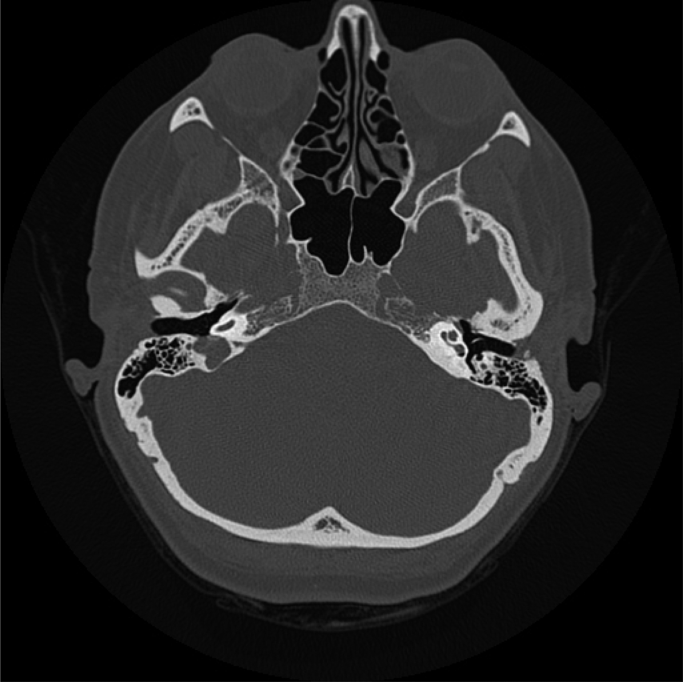
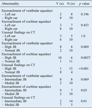
Encroachment of the cochlear aqueduct (Figure 1) by the jugular bulb was apparent in 11 out of 28 affected ears (39.3 per cent), while encroachment of the vestibular aqueduct was apparent in 10 (35.7 per cent). Unusual findings, including diverticulum and dehiscence were observed in eight affected ears (28.6 per cent). There was no significant difference in the incidence of encroachment of either the vestibular or cochlear aqueduct as a function of which ear was affected (p = 0.19 and p = 0.45, respectively; Table III). That is, encroachment of these aqueducts was equally likely to occur in the left and right ears. Similarly, no significant differences were observed in the incidence of unusual CT findings as a function of disease sidedness (p = 1.0; Table III).
Axial computed tomography scan depicting encroachment of the right cochlear aqueduct
Relationship between the position of the jugular bulb and its interaction with surrounding structures
Y = yes; N = no; JB = jugular bulb
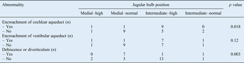
Patients with a high jugular bulb did not have a significantly higher incidence of vestibular aqueduct encroachment by the jugular bulb (p = 0.07; Table III) than those with jugular bulbs considered to be in a normal position. However, encroachment of the cochlear aqueduct was associated with the presence of a high jugular bulb (p = 0.003; Table III). Furthermore, jugular bulb diverticulum or dehiscence was more common in patients whose jugular bulbs were not judged to be high (p < 0.001; Table III). Importantly, the mediolateral position of the jugular bulb also appeared to play a role in encroachment of these structures (Table IV). Further investigation revealed that both intermediate and high jugular bulb positions are associated with encroachment of the cochlear aqueduct, while a medial or normal position is associated with other jugular bulb abnormalities (Table IV).
Mediolateral–anteroposterior positions associated with jugular bulb abnormalities and involvement with surrounding structures
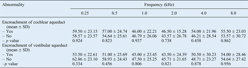
Audiography data was compared in patients who did and did not exhibit encroachment of either the cochlear or vestibular aqueduct. Fisher's exact test indicated no significant difference in audiogram results at any frequency between patients who did and did not exhibit encroachment of the cochlear aqueduct. Similar findings were obtained when comparing patients who did and did not exhibit encroachment of the vestibular aqueduct (Table V).
Hearing loss in patients with and without encroachment of the cochlear and vestibular aqueducts
Discussion
Many studies suggest an atypical location of the jugular bulb to be a possible causative factor in the development of audiological and vestibular disturbances.Reference Friedmann, Eubig, Winata, Pramanik, Merchant and Lalwani2, Reference Wadin, Thomander and Wilbrand8, Reference Couloigner, Grayeli, Bouccara, Julien and Sterkers11, Reference Friedmann, Le, Pramanik and Lalwani18 Other abnormalities of the jugular bulb, such as diverticulum and dehiscence, have also been associated with Ménière's disease-like symptoms.Reference Bilgen, Kirazli, Ogut and Totan6, Reference Hourani, Carey and Yousem7, Reference Schmerber, Lefournier, Lavieille and Boubagra12 However, little is understood about how the anatomical relationship between the jugular bulb and other features of the inner ear might result in such symptoms. Although many hypotheses have been postulated to explain the pathophysiology of Ménière's disease, neither its true cause nor a cure has yet been elucidated.
In this retrospective review of audiometric and radiological data from patients with definite Ménière's disease, the severity of hearing loss was not found to be related to the sidedness of the disease. However, patients with right-sided disease lost hearing at all frequencies compared with their unaffected ears. In contrast, patients whose symptoms occurred in the left ear only did not have significant hearing loss at 4.0 and 8.0 kHz in the affected ear compared with the right ear.
In our population of patients diagnosed with definite Ménière's disease, the observed incidence of high jugular bulb (57.1 per cent) is greater than expected based on previous studies.Reference Friedmann, Eubig, Winata, Pramanik, Merchant and Lalwani1, Reference Vachata, Petrovicky and Sames5, Reference Wadin, Thomander and Wilbrand8–Reference Kuhn, Friedmann, Winata, Eubig, Pramanik and Kveton10 A high jugular bulb is reported to be more common in the right ear.Reference Vachata, Petrovicky and Sames5 Although most of our patients were affected on the right side, our data showed that high jugular bulb did not occur significantly more often in the right ear. Furthermore, high jugular bulb did not occur more frequently in the affected side than in the contralateral ear, suggesting that a high jugular bulb is not the only factor responsible for the development of Ménière's symptoms.
Further analysis indicated that the mediolateral and anteroposterior dimensions of the jugular bulbs in these patients are significantly larger on the affected side than the contralateral side. Moreover, the mean area of the jugular bulbs was significantly greater in affected ears than in unaffected ears, which supports previous reports that the jugular bulb is larger in patients with abnormalities than in those without identified abnormalities.Reference Friedmann, Eubig, Winata, Pramanik, Merchant and Lalwani1
The incidence of encroachment upon the cochlear and vestibular aqueduct was surprisingly high in our study population, suggesting that these encroachments could be related to the disease process. Our results indicate that encroachment upon either of these aqueducts by the jugular bulb is equally likely to occur in the right and left ears. Further investigation suggested that both the anteroposterior and mediolateral positions of the jugular bulb affect encroachment upon the cochlear aqueduct. Thus, the height of the jugular bulb is not the only factor involved in its effects on surrounding structures. Indeed, a high jugular bulb has been found in many patients who exhibit no symptoms. Only 3 out of 26 patients in our study presented with no abnormalities of the jugular bulb. Surprisingly, those without a high jugular bulb were more likely to have dehiscence or diverticulum. This result suggests that the presence of any jugular bulb abnormality may contribute to the development of Ménière's symptoms, presumably by interfering with neighbouring inner-ear structures.
This study has several limitations, including the small number of patients included. Additionally, selection bias may have affected our results because many of the included patients had definite Ménière's disease and had failed medical therapy prior to electing for surgical therapy. Moreover, we did not use a control group of healthy individuals. However, using patients' unaffected contralateral ears as controls may actually be a strength of our study. Directly comparing abnormal and normal jugular bulb anatomy within individuals excludes the potential confounding effects of inter-individual variation in the normal position of the jugular bulb.
Surgical interventions to reposition the jugular bulb have proven useful in treating Ménière's disease-like symptoms, supporting the hypothesis that the jugular bulb may play a role in the development of these symptoms. Studies have reported that the jugular bulb affects Ménière's disease symptoms by obstructing the vestibular aqueduct or, more commonly, the endolymphatic sac or duct, thus preventing drainage of endolymphatic fluids.Reference Bilgen, Kirazli, Ogut and Totan6, Reference Hourani, Carey and Yousem7, Reference Schmerber, Lefournier, Lavieille and Boubagra12, Reference Yetiser, Kertmen and Yildirim21 However, Kawano et al. found no hydrops or otological symptoms in patients whose jugular bulbs were involved with the endolymphatic sac.Reference Kawano, Tono, Schachern, Paparella and Komune22 Experimental studies in guinea pigs demonstrated that endolymphatic–perilymphatic fluid pressure gradients, which are regulated by the cochlear aqueduct, are affected by the presence of hydrops. Furthermore, wide (sac-vein) decompression of the endolymphatic sac, from the sinodural angle to the jugular bulb, has been shown to alleviate Ménière's disease symptoms more effectively than simple endolymphatic sac decompression, suggesting that the endolymphatic sac is not solely responsible for Ménière's disease symptoms.Reference Gianoli, Larouere, Kartush and Wayman23 These results, in conjunction with our data, suggest that the cochlear aqueduct may play a role in the development of Ménière's disease, especially in patients with jugular bulb abnormalities.
• Jugular bulb abnormalities, particularly high jugular bulb, have been implicated in producing Ménière's-like symptoms
• Jugular bulb abnormalities were higher than expected in our patient group
• High jugular bulb was present in 57.1 per cent of affected ears
• Jugular bulb abnormalities were also common in the contralateral ears of Ménière's disease patients
• Mediolateral and anteroposterior jugular bulb positions determine its interaction with surrounding structures
• Cochlear aqueduct encroachment was associated with high jugular bulb
• Abnormal jugular bulb position may contribute to Ménière's disease development
Conclusion
Jugular bulb abnormalities may contribute to Ménière's disease, specifically when the jugular bulb encroaches upon the cochlear aqueduct because this is expected to increase fluid pressure in the inner ear. Cochlear aqueduct encroachment was associated with a high jugular bulb in our study cohort. We do not believe this is the sole cause of Ménière's disease because jugular bulb interference with the vestibular aqueduct and endolymphatic sac or duct is also expected to contribute to the disease. However, the size and position of the jugular bulb may serve as an additional diagnostic marker of disease. The presence of jugular bulb abnormalities may help to identify individuals predisposed to developing Ménière's disease. Because jugular bulb abnormalities are acquired over time and increase blood flow may continue to affect its size and location, jugular bulb abnormalities should be monitored with changing symptomatology.