Introduction
Lymphangiomas are rare, congenital malformations of the lymphatic system. They most commonly occur in the head and neck,Reference Orvidas and Kasperbauer1 with 90 per cent presenting before two years of age.Reference Orvidas and Kasperbauer1 Presentation in adulthood is rare, with approximately 100 cases having been identified internationally.Reference Sherman and Kendall2 Whilst surgical resection remains the recommended treatment, a variety of non-surgical therapies (aspiration, injection of sclerosing agents, diathermy, radiation and observation) have been attempted in children, with varying success rates.
First used in 1987,Reference Ogita, Tsuto, Tokiwa and Takahashi3 the intracystic injection of OK-432, a lyophilised mixture of a low-virulence strain of group A Streptococcus pyogenes incubated with benzylpenicillin, is the primary therapy for lymphangiomatous lesions in Japan. Studies from JapanReference Ogita, Tsuto, Deguchi, Tokiwa, Nagashima and Iwai4, Reference Ogita, Tsuto, Nakamura, Deguchi and Iwai5 and the United StatesReference Greinwald, Burke, Sato, Poust, Kimura and Bauman6–Reference Giguere, Bauman and Smith8 support the effectiveness of this treatment in reducing the size of lymphangiomas, particularly of the macrocystic type. Whilst this therapy is used frequently in children, its effectiveness in adults is unknown, with only two reports of such use appearing in the medical literature.Reference Alonso, Barbier, Alvarez, Romo, Martin and Artteagoitia9, Reference Sichel, Udassin, Gozal, Koplewitz, Dano and Eliashar10
We report an adult case of cystic lymphangioma successfully treated with OK-432, and we review the literature on OK-432 therapy.
Case report
A 31-year-old woman presented to the ENT out-patient clinic with a four-week history of a left-sided neck lump. The lump had appeared spontaneously four weeks after the normal, vaginal delivery of her first child. The lump was not inflamed or painful and the patient was systemically well. She had no past medical history and was taking no medication. She had no known allergies.
On examination, the patient had an obvious cystic lesion, which transilluminated, in the left supraclavicular fossa, lying above the middle third of the clavicle. The lesion was non-pulsatile and had no apparent solid component, and all overlying structures were mobile. The ENT examination, including the thyroid gland, was unremarkable, and there was no cervical lymphadenopathy. Nasendoscopy was normal.
An ultrasound (US) scan demonstrated a simple cyst measuring 5 cm in diameter. Aspiration drained 40 ml of fluid. Cytological analysis demonstrated nuclear pleomorphism, raising the possibility of a lymphoproliferative disorder.
The patient was admitted for formal removal of the cystic lesion under general anaesthesia. The cyst was drained and partially excised. Histological analysis demonstrated a cystic lymphangioma.
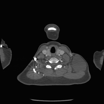
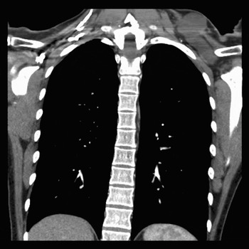
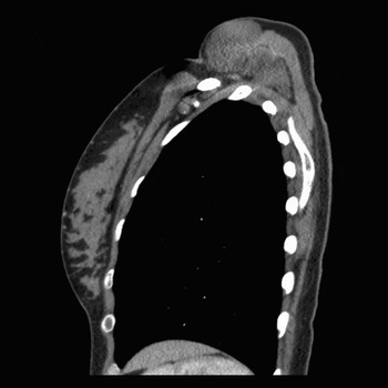
The patient re-attended six weeks later, following a rapid increase in the size of the lesion. Aspiration drained 43 ml of fluid. A computerised tomography (CT) scan was arranged, which demonstrated a cystic lesion tracking behind the trapezius muscle (Figures 1, 2 and 3). Surgery was deemed too difficult, and the patient was thus admitted for injection of OK-432 (Picibanil®; Chugai Pharmaceutical Co. Ltd., Tokyo, Japan) under local anaesthetic. Sixty millilitres of fluid was drained from the cyst and 10 mls of OK-432 was injected.
Axial computed tomography scan of the root of the neck, demonstrating the lymphangioma extending behind the trapezius.

Coronal computed tomography demonstrating extent of the lesion.

Sagittal computed tomography demonstrating extension of the lesion to the level of the scapula.

Post-operatively, the patient developed a rapid increase in the size of the cystic lesion, with an associated inflammatory response and fever. This resolved over five days, and the cyst had disappeared completely six weeks later. Ten months later, there was no recurrence of the lesion.
Discussion
Cystic hygromas are rare tumours, found primarily in children, with the literature describing approximately 100 cases in adults.Reference Sherman and Kendall2 Although most commonly due to abnormal proliferation of lymphoid tissue and thus viewed as a congenital lesion, presentation in adults has been described following trauma, infection, malignancy and iatrogenic stimuli.Reference Orvidas and Kasperbauer1
First classified in 1956,Reference Landing and Farber11 cystic hygromas are commonly divided into three groups: (1) lymphangioma simplex or capillary lymphangioma, (2) cavernous lymphangioma, and (3) cystic lymphangioma or cystic hygroma. A newer classification,Reference Mulliken, Young and Mulliken12 based on the anatomical lesion, histology and CT results, proposes two types of malformations: type one malformations are found below the mylohyoid, are usually cystic without infiltration and are sharply demarcated on CT scan; type two malformations, in contrast, are found above the mylohyoid, usually involve the lip, tongue or oral cavity, are poorly demarcated on CT scan, and demonstrate infiltrative growth patterns with smaller lymphatic channels.
Histologically, the cystic spaces are lined by a single layer of flattened epithelial cells with collections of lymphocytes found in the intervening stroma. The lesions may surround and destroy normal tissue, with the traversing septae often containing thrombosed vessels or degenerate muscle remnants.Reference Sherman and Kendall2
In adults, these lesions usually present as a painless swelling in the neck, with 56 per cent occurring in the posterior triangle and 44 per cent in the anterior triangle.Reference Schefter, Olsen and Gaffey13 There is often no precipitating event, although rapid expansion has been seen following trauma and infection.Reference Orvidas and Kasperbauer1 Although airway obstruction and dysphagia are common presentations of this condition in childhood, they are rare in adults.Reference Alonso, Barbier, Alvarez, Romo, Martin and Artteagoitia9
Relevant investigations include US,Reference El-Silimy and Corney14 CT and magnetic resonance imaging (MRI). T2-weighted MRI is probably superior as it provides high definition scans of neighbouring structures and can assist in planning surgery.Reference Curran, Malik, McShane and Timon15 Aspiration usually reveals straw-coloured fluid.Reference Sherman and Kendall2 The differential diagnosis includes branchial cleft cyst, haemangioma, thyroglossal duct cyst, lymphoma, hamartoma, teratoma, metastatic disease, dermoid cyst, thymic cyst, laryngocoele, thyroid mass and lipoma.
Surgery remains the preferred option for the treatment of cystic hygroma.Reference Orvidas and Kasperbauer1 However, surgery may be technically challenging, particularly if there is intra-thoracic extension. Complications of surgery (including damage to surrounding structures, infection, fistula formation, scarring and recurrence due to incomplete excision) are not infrequent, occurring in 19–33 per cent of patients.Reference Smith, Burke, Sato, Poust, Kimura and Bauman7 Recurrence is related to histology and site of presentation rather than to tumour size or the patient's age.Reference Ricciardelli and Richardson16 Lesions in the suprahyoid region are more likely to cause morbidity, recur and have associated complications, compared with infrahyoid lesions.Reference Ricciardelli and Richardson16
A number of non-surgical therapies have been used, with variable success rates. Radiotherapy, using irradiation or radon-seed implantation, appears to be successful,Reference Martin17 but the risks associated with such treatment limit its use in children to cases of recurrent or persistent, symptomatic disease. Laser therapy, both argon beam and CO2, is effective in debulking or excising lymphangiomas. However, the rate of recurrence is high, necessitating multiple treatments.Reference Giguere, Bauman and Smith8
Aspiration alone is rarely successful unless infection occurs as a complication, causing the lesion to sclerose.Reference Riechelmann, Muehlfay and Teck18 Various sclerosing agents have been used in an attempt to avoid surgery: 50 per cent dextrose,Reference Hancock, StVil, Di Lorenzo and Blanchard19 steroids,Reference Farmaud and Kuttenberger20 alcohol,Reference Stein, Hsu, Schneider, Ruebner and Mina21 Ethibloc®, Reference Dubois, Garel, Abela, Laberge and Yazbeck22 bleomycin,Reference Yura23 cyclophosphamide,Reference Turner and Gross24 fibrin,Reference Castanon, Margarit, Carrasco, Vancells, Albert and Morales25 interferon alfa-2aReference Reinhardt, Nelson, Sencer, Bostrom, Kurachek and Nesbit26 and OK-432.
Alcohol, used for over 100 years in the treatment of varicose veins, is the most potent of all sclerotherapy agents. Used as a 95 or 100 per cent solution, it is cheap and readily available. It acts by damaging the vascular endothelium and denaturing proteins, resulting in a thrombotic reaction. Complications include pain, swelling and bruising. Acute blistering is seen in approximately 25 per cent of patients, and skin necrosis with ulceration occurs in about 21 per cent of patients. This is due either to reflux from superficial venous channels into capillaries, or to alcohol leaking out into the surrounding tissues.Reference Villavicencio27
Ethibloc®, first licensed in West Germany in 1979, is a mixture of corn protein, sodium trizoic, olium papaveris and propylene glycol. It has been administered by percutaneous embolisation to patients with lymphangioma, and its mechanism of action is intravascular necrosis and fibrosis. Although the reported results are good,Reference Dubois, Garel, Abela, Laberge and Yazbeck22 the associated risks include inflammatory reactions and skin ulceration. In addition, Ethibloc may leak through the skin for several months after the injection. Although resolution is usually spontaneous, fistula formation may be unacceptable to many patients.
Bleomycin, an antibiotic with antitumoral activity which is commonly used in chemotherapy regimes, was initially used as a sclerosing agent in 1977,Reference Yura23 with studies reporting excellent clinical results.Reference Orford, Barker, Thonell, King and Murphy28, Reference Tanaka, Inomata, Utsunomiya, Uemoto, Asonuma and Katayama29 The common complications of sclerotherapy with bleomycin include pain, swelling, fever, diarrhoea and vomiting. The most serious complication is pulmonary fibrosis, and, although dose-related, many feel this is an unacceptable risk when the disease being treated is essentially benign.Reference Giguere, Bauman and Smith8
• Cystic lymphangiomas are rare in adults
• Surgery may be technically difficult because of the preponderance of these lesions to extend into the thorax
• OK-432 produces complete regression in children and should be considered as a first line therapy for adults presenting with this condition
Reports of the use of fibrin sealant (Tissucol®) are promising. This haemostatic agent can seal tissue surfaces and eliminate cystic spaces. No complications have been reported, but the number treated is small.Reference Gutierrez, Barrios, Lluna, Menor, Poquet and Ruiz30
The use of interferon alfa-2a to treat lymphangiomas was prompted by its success in treating haemangiomas,Reference Ezekowitz, Mulliken and Folkman31 and the mixed nature of lymphangiomas. Although some benefit has been demonstrated in a small group of children,Reference Reinhardt, Nelson, Sencer, Bostrom, Kurachek and Nesbit26 the general results and the extensive side effects (fever, nausea, diarrhoea, fatigue, alopecia, neutropenia and raised liver enzymes) do not suggest that this treatment should be used widely in the treatment of this condition.
Compared with these other substances, OK-432 produces the most consistent results, and is now recommended by many authors as the treatment of choice for macrocystic lymphangiomas.Reference Giguere, Bauman and Smith8 In 1966, whilst experimenting with anti-cancer therapy, Okamoto discovered that a suspension of avirulent streptococcus (Su) in penicillin G inhibited the growth of malignant ascites in vivo.Reference Okamoto32 Subsequently, this suspension was lyophilised and named OK-432.
Whilst OK-432 lacks the ability to synthesise fatty acids, proteins and nucleic acids and to produce streptolysin-O and -S, the glycolytic activity and some enzymic properties remain.Reference Ishida and Hoshino33
It has been found that OK-432 activates lymphocytes, macrophages and neutrophil killer cells, which destroy tumour cells, and induces cytokines, particularly Th1 cytokines.Reference Ishida and Hoshino33 The interaction between these activated cells and cytokines eliminates tumour cells and accumulated fluid.Reference Ishida and Hoshino33 When OK-432 is injected into cystic hygromas, there is an increase in the production of inflammatory cells, an increase in natural killer cells (CD56+) and T cells (CD3+), and an increase in tumour necrosis factor and interleukin-6.Reference Ishida and Hoshino33 The inflammatory response generated results in shrinkage of the cystic lymphatic spaces, with preservation of the endothelium and no scar formation.Reference Ishida and Hoshino33
Intracystic injection of OK-432 was first reported in Japan in 1987.Reference Ogita, Tsuto, Tokiwa and Takahashi3 Subsequent reports from JapanReference Ogita, Tsuto, Deguchi, Tokiwa, Nagashima and Iwai4, Reference Ogita, Tsuto, Nakamura, Deguchi and Iwai5 and the United StatesReference Greinwald, Burke, Sato, Poust, Kimura and Bauman6–Reference Giguere, Bauman and Smith8 supported the effectiveness of this treatment in reducing the size of lymphangiomas. The treatment appears most suitable for macrocystic lymphangiomas involving the infratemporal fossa or cervical region.Reference Giguere, Bauman, Sato, Burke, Greinwald and Pransky34 Although initially suggested as treatment for lesions not amenable to surgery, reports suggest that OK-432 can also be used as first line treatment.Reference Schmidt, Schimpl and Hollwarth35, Reference Luzzatto, Midrio, Tchaprassian and Guglielmi36
While OK-432 is not licensed for use in the UK, it can be obtained on a named patient basis via IDIS World Medicines. Centres in the UK experienced in the use of this drug include the Great Ormond Street Hospital (London), Royal Liverpool Children's National Health Service Trust, Edinburgh Hospital for Sick Children, Victoria Royal Infirmary (Newcastle upon Tyne), Sheffield Children's Hospital and Frenchay Hospital (Bristol).
The treatment is administered under a general anaesthetic. Contraindications to administration include a history of penicillin allergy or a history of a previous reaction to the product. The manufacturer recommends skin-testing patients with diluted benzylpenicillin before administering OK-432. The OK-432 solution is prepared by dissolving 0.1 mg of OK-432 in 10 ml of normal saline solution (concentration 0.01 mg/ml). A 20-gauge angiocatheter needle is introduced into the cyst. The cyst contents are aspirated through the angiocatheter and the OK-432 solution is then injected into the cyst.Reference Giguere, Bauman, Sato, Burke, Greinwald and Pransky34 Single or multiple cysts can be injected to a maximum total dose of 20 ml (0.2 mg Picibanil). There are no specific guidelines on the desired time space between injections: Smith et al. Reference Smith, Burke, Sato, Poust, Kimura and Bauman7 and Greinwald et al. Reference Greinwald, Burke, Sato, Poust, Kimura and Bauman6 suggest two weeks; OgitaReference Ogita, Tsuto, Deguchi, Tokiwa, Nagashima and Iwai4 suggests three to six weeks; Giguere et al. Reference Giguere, Bauman and Smith8 suggest six to eight weeks; Sung et al. Reference Sung, Lee, Kim, Lee, Hwang and Park37 suggest two to three weeks (unless the mass takes more than one month to shrink; in such cases, a two-month interval is suggested); and Sichel et al. Reference Sichel, Udassin, Gozal, Koplewitz, Dano and Eliashar10 propose waiting three months if progressive tumour shrinkage is seen (see Appendix 1 for suggested management protocol).
Complications include fever, pain and swelling of the area involved.Reference Ishida and Hoshino33 This inflammatory reaction is an important part of the treatment process, and the symptoms usually resolve four days after treatment. Symptomatic management with simple analgesia is usually adequate. Importantly, sclerosis and fibrosis do not occur outside of the cyst, making surgery easier should it be required at a later date.Reference Luzzatto, Midrio, Tchaprassian and Guglielmi36
Studies in children demonstrate complete regression of macrocystic hygromas in 86 to 100 per cent of cases.Reference Ogita, Tsuto, Tokiwa and Takahashi3, Reference Ogita, Tsuto, Nakamura, Deguchi and Iwai5–Reference Smith, Burke, Sato, Poust, Kimura and Bauman7, Reference Giguere, Bauman, Sato, Burke, Greinwald and Pransky34–Reference Laranne, Keski-Nisula, Rautio, Rautiainen and Airaksinen39 Results in microcystic or mixed hygromas are not as promising. Unfortunately, it is difficult to ascertain from the literature how rapidly resolution occurs, but some lymphangiomas may require second and third injections. In adults, there are no large studies as the condition is rare; only two reports of OK-432 use in adults were identified in the literature.Reference Alonso, Barbier, Alvarez, Romo, Martin and Artteagoitia9, Reference Sichel, Udassin, Gozal, Koplewitz, Dano and Eliashar10 However, given its success in children, there appears to be no reason why this treatment should not be used routinely in adults.
Conclusion
Cystic lymphangiomas are rare in adults and may appear following trauma, infection or malignancy, or secondary to an iatrogenic cause. In children, OK-432 injection appears to be the treatment of choice for macrocystic lymphangiomas. This treatment should be considered in adult patients presenting with similar lesions.
Appendix 1. Use of OK-432 (Picibanil®)
1 Identify patient who would potentially benefit from treatment (ideally, a patient with a macrocystic lymphangioma).
2 Ensure that patient does not have a hypersensitivity reaction to penicillin. Perform skin test if necessary.
3 Locate source of drug (IDIS World Medicines; handling charge imposed; maximum of 10 packs for single, named patient use).
4 Treatment is administered under general anaesthetic.
5 Dissolve 0.1 mg of OK-432 in 10 ml of normal saline solution (produces a solution with a concentration of 0.01 mg/ml).
6 Aspirate the contents of the cystic lesion.
7 Inject the OK-432 solution to a maximum dose of 20 mls (0.2 mg OK-432).
8 Side effects include fever, pain and swelling of the area involved. These last four to seven days and should be treated with simple analgesics and anti-pyretics.
9 If necessary, repeat the injection two to eight weeks after the initial injection. Time interval between injections will depend upon the response to initial therapy.
10 Centres with experience in the use of this drug include: Great Ormond Street Hospital, London; Royal Liverpool Children's National Health Service Trust; Edinburgh Hospital for Sick Children; Victoria Royal Infirmary, Newcastle upon Tyne; Sheffield Children's Hospital; and Frenchay Hospital, Bristol.