I. INTRODUCTION
Recent experimental and industrial advances in the field of nanotechnology have boosted the development of interdisciplinary research, one of the most constructive and inspiring human pursuits. Reference Yao, Sun, Yang and Duan1,Reference Yang, Yao and Duan2 Semiconductor quantum dots (QDs), as a kind of increasingly important nanosized materials, have attracted great interests in many fields across the physical, chemical, and biological sciences and become well-established photoluminescent platforms for scientific and industrial applications. Reference Kamat, Tvrdy, Baker and Radich3–Reference Samia, Chen and Burda11 Compared with typical organic dyes and fluorescent proteins, semiconductor QDs offer several unique advantages, such as high absorption cross sections and narrow/symmetric emission spectra, which make them highly suitable for using as reporters for multiplexing assays. Reference Shi, Wang, Li, Wang, Zang, Zu, Zhang, Guo and Tong12–Reference Zhou, Zhou, Du, Bi and Zhou16 In addition, QDs exhibit high fluorescence quantum yields, high stability against photobleaching, and a pronounced resistance to photo- and chemical-degradation, endowing them the possibility of continuous or long-term monitoring slow biological processes. Reference Jaiswal, Mattoussi, Mauro and Simon17,Reference Somers, Bawendi and Nocera18 These unique properties of QDs make them appealing as in vivo and in vitro fluorophores in a variety of biological investigations. Reference Dubertret, Skourides, Norris, Noireaux, Brivanlou and Libchaber19–Reference Chen, Zhou, Chang, Ren, Chen and Chen21 The controlled fluorescence quenching of QDs, such as CdTe, CdSe, ZnSe, CdS, and ZnS, has led to the development of probes for sensing a variety of molecules such as enzyme, DNA, nitric oxide, anions, and cations. Reference Qiao, Jian-Hao, Zhan, Zhao-Hui, Qin and Yuan-Di22–Reference Wang, Yang, Fang, Wu, Liu, Min and Li26
Hemin, the oxidized form of iron protoporphyrin IX, is an iron-containing complex and often can be regarded as an essential regulator of gene expression and growth promoter of hematopoietic progenitor cells. It is usually selected as an oxidant in several systems involving the catalytic and prooxidant action of iron. Reference Tsiftsoglou, Tsamadou and Papadopoulou27–Reference Lu, Zhang, Li and Gao29 Nowadays, the analysis of hemin mainly adopts electrochemical method, fluorescence, and chemiluminescence. Among these, fluorescence is the most effective technique because it owns high sensitivity and excellent response time. However, there are little papers that study the fluorescence behavior of hemin and QDs, especially the CdTe system.
Here, we described a fast and eco-friendly method to successfully prepare highly luminescent CdTe QDs in an aqueous solution. The obtained QDs were characterized by transmission electron microscopy (TEM), selected area electron diffraction (SAED), scanning transmission electron microscope–energy dispersive x-ray spectroscopy (STEM-EDX), Fourier transform infrared (FTIR), UV-vis and photoluminescence (PL) spectroscopy, respectively. Then, we demonstrated the use of CdTe QDs to optically detect hemin based on the fluorescence quenching of QDs. This method was sensitive and provided a wide linear range of hemin concentrations under the optimal conditions. We also investigated the detection mechanism of hemin and found that the quenching effect was caused by the electron transfer from hemin to QDs.
II. EXPERIMENTAL
A. Chemicals
3-Mercaptopropionic acid (MPA) and hemin were purchased from Aladdin (Shanghai, China); NaBH4 and CdCl2·2·5H2O were purchased from Kelong Chemical Company (Chengdu, China); Tellurium powder was purchased from Shanghai Chemical Reagent Company (Shanghai, China). All other reagents were of analytical reagent grade and used without further purification. Ultrapure water was used throughout the experiments.
B. Synthesis
CdTe QDs in a MPA-capped form were synthesized as follows. First, sodium hydrogen telluride (NaHTe) was prepared by reducing Te powder with sodium borohydride in ultrapure water under stirring and N2 purging. Then, CdCl2 was dissolved in water in a flask followed by the addition of pure MPA under vigorous stirring. After MPA was completely dissolved, the pH of the solution was adjusted by dropwise addition of NaOH solution. In the next step, the fresh NaHTe was injected into N2-saturated CdCl2 mixture, and the resulting crude solution was refluxed for a certain time to promote the growth of CdTe QDs.
C. Characterization
1. Optical characterization
Optical absorption spectra of CdTe QDs were recorded with a Genesys 10S UV-vis spectrophotometer (Thermo Fisher Scientific Co., Madison, WI), using a 1.0 cm-quartz cuvette. Fluorescence spectra were recorded on a Perkin–Elmer LS-55 fluorescence spectrometer (Perkin-Elmer Ltd., Llantrisant, Wales, UK). All optical measurements were performed under ambient conditions.
2. TEM, SAED, and STEM-EDX characterization
TEM and SAED images of the NPs were acquired on a Tecnai G2 F20 TEM (FEI Company, Hillsboro, OR). In this case, one drop of CdTe QDs was placed onto a holey carbon 400 mesh TEM grid and was allowed to evaporate under ambient conditions. STEM-EDX images were operated at an acceleration voltage of 200 kV.
3. FTIR spectroscopy
FTIR spectral analysis was done using a Nicolet 6700 spectrometer (Thermo Fisher Scientific Co., Madison, WI).
III. RESULTS AND DISCUSSION
A. Optical properties of MPA–CdTe
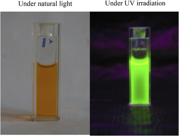
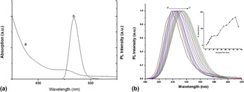
A digital camera was used to observe the solution of MPA–CdTe in deionized water under visible light and ultraviolet light excitation in a darkroom. The photographs were presented in Fig. 1. The solution exhibited orange colors under visible light, while after being radiated under a hand-held UV lamp, it emitted green fluorescence. Then, the as-prepared CdTe QDs solution was further characterized using a UV-vis scanning spectrophotometer and a fluorescence spectrophotometer, respectively. The UV-vis absorption and room temperature FL spectra of CdTe QDs were displayed in Fig. 2(a). As shown in curve (a), the obvious UV-vis absorption peak of CdTe QDs was centered at 499 nm. The diameter of QDs was calculated by the wave length of the first excitonic absorption peak of the corresponding sample according to the empirical fitting functions. Reference Yu, Qu, Guo and Peng30 It is estimated to be about 2.3 nm. From curve (b), it can be seen that CdTe QDs have a narrow emission peak: the full width at half-maximum is 40 nm, which shows that the as-prepared CdTe QDs are nearly monodisperse and homogeneous. The maximum of the fluorescence emission appeared at 535 nm. The optical performance of a series of CdTe QDs synthesized with different reaction times was examined, as shown in Fig. 2(b). It shows that the emission peak continuously shifts to longer wave length with prolonging heating time, which corresponds to the increase of particle size. At the same time, the PL intensity first increases and then declines. The PL intensity significantly increased because the surface traps of the CdTe QDs were effectively eliminated by CdS shell, which originated from the decomposition of MPA. As the refluxing time was prolonged, the PL intensity reached a maximum value and then started to decrease; this was possibly due to dislocations and new defects that were formed with the MPA being consumed.
Pictures of MPA-capped CdTe QDs under natural light and UV irradiation.

Representative absorption and PL spectra (a) and temporal evolution of the PL emission spectra (b) of MPA-capped CdTe QDs.

B. Reaction mechanism of MPA
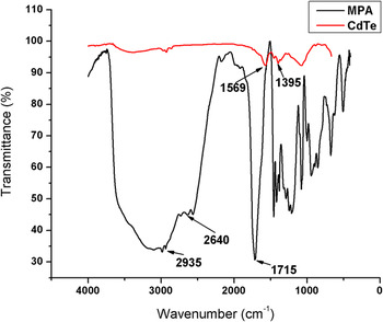
FTIR has been one of the most common methods to determine the molecular interactions between the NPs and their capping agents. The structural features of MPA-stabilized CdTe QDs were investigated by FTIR (Fig. 3). In the FTIR spectrum of MPA, the strong absorption in the range of 3425–3034 cm−1 is assigned to the O–H vibration of MPA multimer. The signal around 2640 and 2935 cm−1 can be attributed to S–CH3 and S–H stretching modes, respectively. Besides that, the peak near to 1715 cm−1 relates to the C=O stretching of COOH. However, as expected, in comparison with the clear S–H stretching peak in MPA, the corresponding peak disappears in the FTIR spectrum of MPA–CdTe, which indicates that the thiol group of MPA is bound to surface atoms of QDs through the Cd–S bond. On the other hand, the appearance of the bands at 1569 and 1395 cm−1, which are attributed to asymmetric νas (COO–) and symmetric νs (COO–) vibrations, respectively, Reference Colthup, Daly and Wiberley31 for the CdTe nanoparticles, indicates that MPA exists in the form of carboxylate salt and reveals that MPA is chemisorbed as a carboxylate onto Cd ions on the CdTe NPs surface. The carboxylic acid is known to be absorbed on the metal as a carboxylate after deprotonation. Reference Han, Han and Kim32,Reference Tao33
FTIR spectra of MPA and MPA–CdTe QDs.

C. Morphology analysis
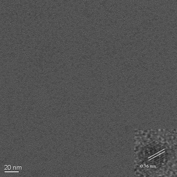
The morphological analysis of the as-synthesized MPA-capped CdTe was carried out using both TEM and high resolution transmission electron microscopy (HRTEM) (Fig. 4). It is quite evident that these nanocrystals are close to sphere with excellent monodispersity. Based on the TEM results, size distribution analysis indicates that the average particle size is around 2.3 nm for CdTe QDs, which is in agreement with the calculated value. The HRTEM image of CdTe QDs (inset) indicates that the synthesized NPs are crystalline in nature and they exhibit well-resolved lattice fringes. The distance between two adjacent planes is estimated to be 0.36 nm, corresponding to the (111) lattice plane of cubic CdTe (JCPDS No. 65-0890).
TEM and HRTEM (inset) images of CdTe QDs.

D. Crystal structure
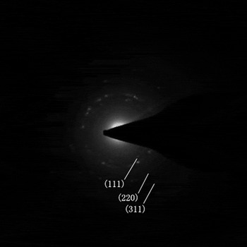
Moreover, to verify the state of existence of CdTe NPs, the SAED patterns were obtained from a selected area of the CdTe QDs via a convergent beam, as shown in Fig. 5. They appear as distinguishable diffraction rings, inferring that the QDs are small nanocrystals. The existence of well-resolved lattice planes in the SAED image further confirms the excellent crystalline structures of the as-prepared CdTe QDs. The distances between the CdTe wafers, d, are 0.363, 0.234, and 0.189 nm, which correspond to the (111), (220), and (311) planes, indicating that the CdTe NPs are cubic lattice. Reference Wang, Yang, Bai, Song, Zhang and Li34,Reference Zhang, Zhou, Yang and Gao35
SAED pattern of CdTe QDs.

E. Semiquantification of MPA–CdTe
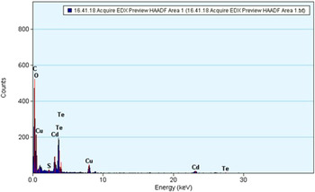
Furthermore, the semiquantitative picture on the surface composition of CdTe NPs was investigated by using STEM-EDX. As displayed in Fig. 6, the STEM-EDX pattern clearly confirms the presence of Cd and Te, and the atomic ratio of Cd:Te calculated from EDX analysis is 1:1.15. The high proportion of C is considered from MPA capped on CdTe, unreacted MPA remained after purification and Cu grid coating, while the O is expected from MPA that bond to CdTe or unreacted MPA, from adventitious atmosphere.
STEM-EDX spectrum of MPA-capped CdTe QDs.

F. Application for biomolecule analysis
1. Optimization of experimental conditions
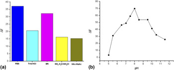
Since the fluorescence of MPA-capped CdTe QDs is very sensitively affected by the kind and pH of buffer solution, the effect of these two important parameters on the response of the sensing material to hemin is studied. Figure 7(a) shows the relative fluorescence intensity of QDs with 1 × 10⁻⁵ mol/L hemin in phosphate buffer solution (PBS), Tris–HCl, Britton–Robinson (BR) buffer, NH3·H2O–NH4Cl, and HAc–NaAc at pH = 7.4, respectively. Clearly, it can be observed from Fig. 7(a) that the fluorescence intensity of CdTe–hemin in the PBS solution is the most remarkably quenched. So, in this paper, PBS was selected to control the pH of system. Investigation of the effect of pH value on the performance of the hemin sensor is of great importance, because the surface of QDs and the interaction between QDs and hemin are pH dependent. Therefore, the effect of pH in a range between 5 and 11.5 was studied to select the optimum conditions for the determination of hemin. In the acidic environment, the fluorescence of QDs is very weak, and this may be explained as that the –SH tends to fall off and lattice defects will exist in QDs, which usually causes the decrease or disappearance of QDs fluorescence. When the pH varies from 7 to 11.5, the QDs fluorescence is strong enough to act as a very good signal display unit. In addition, the influence of pH on the interaction of hemin and QDs was explored. As demonstrated in Fig. 7(b), the quenching effect of hemin is first enhanced and then decreased with increasing pH. The maximum quenching effect can be obtained at pH 8.0, maintained by 0.1 M phosphate buffer.
Effect of (a) buffer solutions and (b) pH on the interaction between QDs and hemin.

2. Efficient quenching effect of hemin on QDs PL intensity
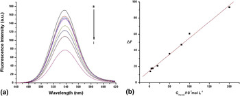
Under the optimum conditions, the fluorescence emission intensity of the assay was measured after the addition of various amounts of hemin recorded at pH 8.0 (as a representative of the rest of the pH value). A dose–response graph was obtained to determine the relationship between the fluorescence intensity of CdTe QDs and the concentration of the quenching hemin. Figure 8 shows quenching effects of hemin with different concentrations on the fluorescence of QDs. As can be seen, with the addition of hemin to the MPA-capped CdTe QDs aqueous solution, the fluorescence intensity continuously decreased and no visible shape changes of fluorescence spectra were observed. The calibration curve was prepared by adding a series of hemin standard solutions at different concentrations and the correlation of hemin concentration with the relative fluorescence intensity provided the basis for quantitative analysis. The fitted curve showed a good linear relationship between ΔF and hemin concentration within a range of 2.0 × 10⁻⁷–2.0 × 10⁻⁵ M. It was found that a relatively broad linear range could be obtained. The linear regression equation is described by the following equation: ΔF = 0.4058 C + 14.7893 (ΔF = F 0 − F, where F 0 and F represent the fluorescence intensity without and with hemin, C is the concentration of hemin). The correlation coefficient and limit of detection (LOD) at 3S 0/S are 0.9960 and 8 × 10⁻⁸ M, respectively. Therefore, the observed quenching effect can be used to monitor the concentration of hemin.
(a) Fluorescence spectra of MPA–CdTe in the presence of different concentrations of hemin. The concentrations of hemin were 0 (a), 2 × 10⁻⁷ (b), 5 × 10⁻⁷ (c), 8 × 10⁻⁷ (d), 2 × 10⁻⁶ (e), 5 × 10⁻⁶ (f), 8 × 10⁻⁶ (g), 1 × 10⁻⁵ (h), and 2 × 10⁻⁵ (i); (b) plot of relative fluorescence intensity versus hemin concentration.

3. Study on the quenching mechanism of QDs by hemin
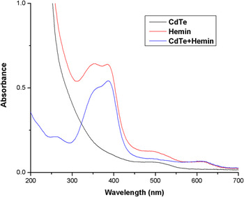
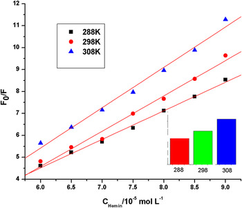
Generally, the quenching of the fluorescence emission originates either from static or dynamic interaction of quencher with the fluorophore. Static quenching refers to the formation of a complex between quencher and fluorophore, which cannot emit a photon. Dynamic quenching is known to occur mainly by an intermolecular collisional process. Therefore, UV-vis absorption measurement is a very facile method to investigate the fluorescence quenching mechanism between fluorophore and quencher. Figure 9 presents the absorption spectra of CdTe, hemin, and CdTe–hemin mixture. For hemin, there are four absorption peaks at 270, 350, 390, and 620 nm, respectively, whereas CdTe QDs only show one peak at 499 nm. From Fig. 9, we can see that the absorption spectrum of CdTe–hemin system is the superposition of hemin and CdTe. No new band or peak shift is observed in the absorption spectrum of CdTe–hemin system, which indicates that the quenching of CdTe QDs may not be caused by the structural changes or the complex formation (static quenching). Reference Huang, Geng, Liu, Wang, Jiao and Yu36–Reference Mikhail and Achilefu41 It is perhaps responsible for a collisional process (dynamic quenching). The quenching of QDs fluorescence by hemin can be described by the well-known Stern–Volmer equation Reference Lakowicz42 : F 0/F = 1 + K qτ0[Q] = 1 + K sv[Q], where F 0 and F denote the fluorescence intensities of MPA–CdTe at λem when excited at λex in the absence and presence of quencher, respectively; K q is the quenching rate constant; τ0 is the average fluorescence lifetime of the system in the absence of quencher; [Q] is the concentration of quencher; and K sv is the Stern–Volmer fluorescence quenching constant. Hence, the above equation could be applied to determine K sv by linear regression of a plot of F 0/F versus [Q]. Figure 10 shows the typical Stern–Volmer plots for CdTe–hemin system at three different temperatures. As seen, the quenching constants increased with increasing temperatures. As depicted in the inset of Fig. 10, K sv of the MPA–CdTe QDs–hemin system at three temperatures was calculated according to the Stern–Volmer equation. It can be seen that the quenching type of MPA–CdTe QDs–hemin system is dynamic quenching.
Absorption spectra of CdTe, hemin, and CdTe–hemin system.

Stern–Volmer plots for the quenching of QDs by hemin at different temperatures. The inset shows the relationship of the Stern–Volmer quenching constants K sv and temperature.

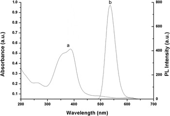
Plenty of studies point out that the quenching of QD fluorescence can be ascribed to Förster resonance energy transfer (FRET), adsorption of molecules, and electron transfer. FRET is a process that involves the transfer of fluorescence energy from a donor particle, D, (after absorption of a higher-energy photon) to another proximal ground-state acceptor, A, via a resonant, short range dipole–dipole interaction. Reference Azzazy, Mansour and Kazmierczak43–Reference Pons and Mattoussi45 To clarify the possibility of FRET, the fluorescence spectrum of MPA-capped CdTe QDs and the absorption spectrum of hemin were recorded. From Fig. 11, we can see that the emission spectrum of QDs does not overlap with the absorption spectrum of hemin. Therefore, the hypothesis of energy transfer is untenable for CdTe–hemin system. After adding hemin in the solution, the fluorescence intensity of CdTe QDs decreased significantly while the absorption spectra did not shift to blue or red, indicating that the quenching of QDs by hemin was not attributed to molecular adsorption. As described in Fig. 8(a), with the change of hemin concentration, there was no shift of the fluorescence emission spectra, demonstrating that CdTe QDs did not aggregate or shrink in the presence of quenchers. Reference Sun, Ren, Liu and Chen46 It is generally known that the fluorescence of QDs originates from the recombination of electron and hole. Hemin as a fluorescence quencher can donate its electron to CdTe QDs to occupy the hole and accordingly disrupt the radioactive recombination process and quench the fluorescence.
UV-vis absorption spectrum of hemin (a) and luminescence spectrum of MPA–CdTe QDs (b).

IV. CONCLUSION
In conclusion, MPA-capped CdTe QDs were synthesized in an aqueous solution through a convenient, one-pot strategy by using safe and low-cost inorganic salts as precursors. Many analytical techniques including but not limited to TEM, HRTEM, STEM-EDX, SAED, PL spectra, UV-vis spectra, and FTIR were used to explore their structural, photochemical, and photophysical properties. It demonstrated that the as-prepared CdTe QDs were near-spherical and monodispersed particles. They are cubic structures with a diameter of 2.3 nm. MPA can stabilize the CdTe surface and endow QDs excellent water solubility and biocompatibility. Moreover, a smart and novel method for investigating the interaction of CdTe QDs–hemin system was proposed. The results indicated that the quenching of QDs fluorescence was a collisional process rather than a static quenching process and electron transfer played a major role in the interaction where hemin quenched the PL of CdTe QDs. In addition, the wide detected range, low LOD, simplicity, and rapidity of this method provide CdTe QDs a promising fluorescence probe for hemin sensing.
ACKNOWLEDGMENTS
The authors are grateful for the financial support from National Major Scientific Instruments and Equipments Development Special Funds (Grant No. 2011YQ030113), National Natural Science Foundation of China (Grant No. 21275105), National Recruitment Program of Global Experts (NRPGE), the Hundred Talents Program of Sichuan Province (HTPSP), and the Startup Funding of Sichuan University for setting up the Research Center of Analytical Instrumentation.