Case report
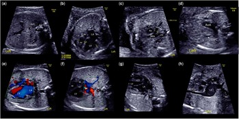
A 29-year-old woman, gravida 2, para 1, with a dichorionic–diamniotic twin pregnancy, was referred at 20 weeks of gestation for prenatal echocardiography because of a heart defect detected in one fetus. Prenatal echocardiography revealed severe heart asymmetry related to isolated total anomalous systemic venous drainage to the left atrium (Fig 1). Right-sided and left-sided superior caval veins, the inferior caval vein, and the hepatic veins were all connected to the left atrium. All pulmonary veins drained normally into the left atrium. The interatrial septum appeared thickened with a restrictive foramen ovale. The ventricles were unbalanced, with a smaller right ventricular chamber compared with the left. Despite this significant ventricular asymmetry, the main pulmonary artery was relatively well developed, with antegrade flow from the right ventricle to the pulmonary trunk. In addition, pulmonary perfusion was partially provided by the arterial duct, which exhibited bidirectional flow.
Fetal echocardiography at 23 ( a – c ) and 27 weeks ( d–h ) of gestation. ( a ) Abdominal axial view showing the inferior caval vein (IVC) located anterior to and to the right of the descending aorta (Ao). The IVC is crossing the midline closer to the diaphragm to join the left atrium (LA) (not shown). ( b ) Four-chamber view showing the heart asymmetry. Of note, the right ventricular (RV) length is quite well preserved, despite its abnormally small diameter. ( c ) Axial view at the level of the “three-vessel and trachea view” demonstrating the presence of an additional vein on the left side of the main pulmonary artery (MPA): a left superior caval vein (LSVC). ( d ) By moving the transducer slightly more cranially, we were able to reveal the right superior caval vein (RSVC) and LSVC as well as the left aortic arch (AoA). ( e ) Four-chamber view with color Doppler imaging showing a small patent foramen ovale (FO) with left-to-right shunting and two patent but asymmetric atrioventricular valves. ( f ) Four-chamber view with color Doppler imaging demonstrating normal right and left pulmonary venous connection to the LA. ( g ) Four-chamber view showing the drainage of the RSVC and LSCV directly into the LA. ( h ) Sagittal view demonstrating the drainage of the RSVC and IVC into the LA posterior to the right atrium (RA). LPA=left pulmonary artery; LPV=left pulmonary vein; LV=left ventricle; RPA=right pulmonary artery; RPV=right pulmonary vein.

At serial follow-up, the right heart structures were observed to still be growing, with slightly milder asymmetry seen over time. The tricuspid-to-mitral valve annulus and main pulmonary-to-ascending aorta ratios increased from 0.48 to 0.62 and from 0.68 to 0.76, respectively, from 23 weeks to 33 weeks of gestation.
At 36 weeks of gestation, an emergency caesarean section was performed because of placental abruption. The newborn infant suffering from cardiac malformation adapted well to extrauterine life, exhibiting APGAR scores of 4/7/8 at 1, 5, and 10 minutes and birth weight of 2.2 Kg. Transcutaneous saturation reached 88% with oxygen therapy. Clinical examination revealed no abnormalities or dysmorphic features.
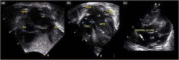
The prenatal diagnosis was confirmed by postnatal echocardiography (Fig 2), and no other cardiac anomaly was associated. Both right and left superior caval veins and the inferior caval vein drained into the left atrium. The left atrium was connected to the morphological left ventricle. The shunt through the restrictive foramen ovale was directed from left to right. The antegrade pulmonary perfusion was insufficient, requiring prostaglandin administration in order to maintain ductal patency. The right atrium was hypoplastic, and no vascular structure drained into the bald, smooth right atrium. The right ventricle was relatively small but tripartite. Pulmonary venous return was normal.
Postnatal echocardiography. ( a ) Subcostal view showing the right superior caval vein (RSVC) and the inferior caval vein (IVC) draining into the left atrium (LA). ( b ) Four-chamber view showing the small “bald”-looking right atrium (RA) and the large LA receiving the RSVC and left superior caval vein (LSVC) and demonstrating the mild asymmetry between the two atrioventricular valves and ventricles. ( c ) The central intravenous line placed into the RSVC is shown entering into the LA. LV=left ventricle; MITR=mitral valve annulus; RV=right ventricle; TRIC=tricuspid valve annulus.

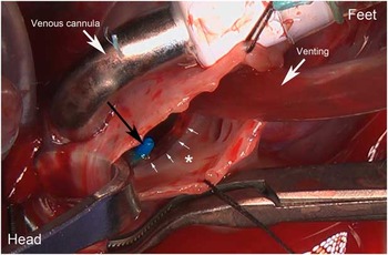
To improve flow to the right heart and enable sufficient pulmonary flow, an atrial septectomy was performed (Fig 3) under cardiopulmonary bypass together with a modified right-sided 3.5mm Blalock–Taussig shunt and arterial duct ligation and section 3 days after birth. Total anomalous systemic venous connection to the left atrium was confirmed during surgery. Surgical findings confirmed a right ventricle of reasonable size that reached the apex of the heart.
Operative view following atrial septectomy. Notice the small right atrium with a smooth surface (*). The small white arrows indicate the posteroinferior rim of the residual atrial septum. The black arrow shows the tip of the central venous line placed in the right superior caval vein entering the left atrium.

Unfortunately, the newborn’s immediate postoperative course was marked by bradycardia, progressive hypotension, and signs of low cardiac output, which were treated by means of intravenous fluid therapy and inotropic drugs. Cardiac arrest occurred 6 hours following the operation, which was followed by unsuccessful cardiopulmonary resuscitation and death. The parents refused autopsy.
Discussion
Congenital anomalies of the systemic venous return are a heterogeneous group of cardiac malformations with widely variable physiological consequences.Reference Mazzucco, Bortolotti, Stellin and Gallucci 1 The most common type is persistence of the left superior caval vein, whereas total anomalous systemic venous connection to the left atrium is a rare entity. Among 100 patients with anomalous systemic venous connections undergoing surgery, only 3 patients have a total anomalous systemic venous connection to the left atrium.Reference de Leval, Ritter, Mc Goon and Danielson 2 Only a few cases have been reported thus far,Reference Zhang, Duan, Gao, Ru and Ying 3 – Reference Vaidyanathan, Kothandam, Kumar, Pradhan and Agarwal 8 and the literature on this subject has more of a surgical perspective. Most patients presented later in life, from childhood to adulthood, mainly with cyanosis. To our knowledge, this is the first case report of a prenatal diagnosis of total anomalous systemic venous connection to the left atrium.
Most cases of total anomalous systemic venous connection to the left atrium reported in the literature were associated with left isomerism.Reference Awasthy, Radhakrishnan, Kaushal and Sharma 5 – Reference Vaidyanathan, Kothandam, Kumar, Pradhan and Agarwal 8 On surgical inspection of the appendage of the right-sided atrium, we found that it exhibited the anatomical characteristics of a left-atrial appendage – namely, a long narrow chamber with a smooth-looking wall, possibly indicating heterotaxy syndrome with left-atrial isomerism. The chest X-ray, however, did not show bilateral symmetric left bronchi with widened carina as typically observed in left bronchial isomerism. No abdominal ultrasound was performed to confirm midline liver and polysplenia.
Total abnormal systemic venous return requires a left-to-right shunt through an atrial septal defect, a ventricular septal defect, or a patent arterial duct in order to ensure pulmonary circulation. A large atrial septal defect was present in all patients of studies previously publishedReference Zhang, Duan, Gao, Ru and Ying 3 – Reference Yildirim, Kosger, Ozdemir, Ucar and Kilic 6 , Reference Vaidyanathan, Kothandam, Kumar, Pradhan and Agarwal 8 except for two of them. A 7-year-old girl had a large perimembranous ventricular septal defect,Reference Vo, Cao, Le and Nguyen 7 and a 2-year-old boy had only a small atrial septal defect.Reference Awasthy, Radhakrishnan, Kaushal and Sharma 5 In the latter case, the right ventricle was hypoplastic and bipartite, and single ventricle palliation was offered with success. In all other patients, biventricular repair has been possible, thanks to good right ventricular development secondary to unrestrictive left-to-right shunt. In our case, shunting through the tiny foramen ovale was insufficient, and the pulmonary arteries were perfused, at least partially, through the arterial duct, which was maintained open by means of prostaglandin infusion immediately after birth.
Owing to a thickened interatrial septum, a fairly small right ventricle, and the low birth weight, we decided to enlarge the foramen ovale by surgery and to perform a modified right-sided Blalock–Taussig shunt at the same time in order to postpone corrective surgery to a later date; yet, the child exhibited low cardiac output shortly after the surgery, along with severe left ventricular dysfunction and pulmonary oedema, finally suffering cardiac arrest. This might have been accounted for by pulmonary overcirculation and sudden volume overload of the left heart.
Despite an unsuccessful postoperative course, this report primarily aims to describe the first prenatal description of this very rare congenital heart malformation.
Acknowledgements
The authors are grateful to their colleagues who referred the patient to them and to those who took care of the child after birth.
Financial Support
This research received no specific grant from any funding agency or from commercial or not-for-profit sectors.
Conflicts of Interest
None.
Ethical Standards
The authors assert that all procedures contributing to this study comply with the ethical standards of the relevant Belgian national guidelines on human experimentation and with the Helsinki Declaration of 1975, as revised in 2008, and has been approved by the institutional committee of the Cliniques universitaires Saint-Luc.