Introduction
Eosinophilic granulomatosis with polyangiitis, formerly known as Churg–Strauss syndrome, is a systemic, necrotising vasculitis characterised by peripheral blood eosinophilia and eosinophilic infiltration of involved tissues.Reference Groh, Pagnoux, Baldini, Bel, Bottero and Cottin1
Eosinophilic granulomatosis with polyangiitis has significant ENT involvement, especially in the nose and paranasal sinuses,Reference Masi, Hunder, Lie, Michel, Bloch and Arend2 with chronic rhinosinusitis with and without nasal polyposis being the most common features.Reference Bacciu, Bacciu, Mercante, Ingegnoli, Grasselli and Vaglio3 Similarly, Wegener's granulomatosis, currently named granulomatosis with polyangiitis, can affect the nose: frequently, these patients are afflicted with nasal deformity, nasal crusting and septal perforations.Reference Gouveris, Karaiskaki, Koutsimpelas, Chongolwatana and Mann4
As far as other ENT sites are concerned, the larynx can be one of the targeted organs in granulomatosis with polyangiitis, with up to 14 per cent of patients affected,Reference Gouveris, Karaiskaki, Koutsimpelas, Chongolwatana and Mann4–Reference Del Pero, Jayne, Chaudhry, Sivasothy and Jani6 but much less is known about laryngeal involvement in eosinophilic granulomatosis with polyangiitis.
A literature review was performed to investigate whether there is a similarity between eosinophilic granulomatosis with polyangiitis and granulomatosis with polyangiitis, in terms of the nose and the larynx. This was accompanied by a prospective cross-sectional study of the eosinophilic granulomatosis with polyangiitis patients who attended the out-patient clinic of our rheumatology unit. In particular, we questioned whether eosinophilic granulomatosis with polyangiitis patients had subglottic stenosis or simple laryngeal inflammation, by means of conventional white light endoscopy coupled with narrow-band imaging. Narrow-band imaging endoscopy uses narrow-bandwidth filters in a sequential red–green–blue illumination system, and can enhance visualisation of mucosal vascular damage in inflamed tissues resulting from epithelial inflammation.Reference Wang and Tsai7
Materials and methods
The clinical research was preceded by a comprehensive literature search, performed using PubMed and Medline databases, with the key words: ‘larynx’, ‘subglottic stenosis’, ‘eosinophilic granulomatosis with polyangiitis’, ‘granulomatosis with polyangiitis’, ‘Churg-Strauss Syndrome’ and ‘Wegener's granulomatosis’.
The literature search was accompanied by a cross-sectional prospective study, which started in 2010, of an unselected group of eosinophilic granulomatosis with polyangiitis patients, who attended the University Hospital of Pisa for regular check-ups. Our hospital is a referral centre for systemic vasculitis cases in Italy. It provides an interdisciplinary approach, involving rheumatologists, otolaryngologists and pulmonologists. All eosinophilic granulomatosis with polyangiitis patients undergo a thorough check-up, so ENT evaluation is performed in every case, even in the absence of specific symptoms.
All patients included in our study were previously diagnosed with eosinophilic granulomatosis with polyangiitis accordingly to American College of Rheumatology criteria.Reference Masi, Hunder, Lie, Michel, Bloch and Arend2 All patients were undergoing therapy with oral steroids and/or disease-modifying drugs, including methotrexate, azathioprine and mycophenolate mofetil. The patients were also receiving proton-pump inhibitors as a preventive treatment for steroid-linked gastropathy.
At the time of the ENT evaluation, the rheumatologist considered the patients’ condition as being under control, based on various disease activity clinical scores, such as the Birmingham Vasculitis Activity ScoreReference Luqmani, Bacon, Moots, Janssen, Pall and Emery8 and Vasculitis Damage Index.Reference Exley, Bacon, Luqmani, Kitas, Gordon and Savage9 The Birmingham Vasculitis Activity Score in particular assesses disease activity, at a given time point, as the sum of individual organ system manifestations caused by active disease, defined by a list of 34 weighted items, with respect to whether symptoms are new, worse or persistent. Along with nasal crusting and sinus involvement, another ENT item specifically assessed during the ENT evaluation was ‘subglottic inflammation’.
In fact, laryngeal endoscopy was performed in all patients, and the status of the vocal folds, and the supraglottic and subglottic regions, were registered. We also recorded the presence of laryngeal inflammation by means of the Reflux Finding Score.Reference Belafsky, Postma and Koufman10 All laryngoscopies, performed under conventional white light, involved a (non-invasive) narrow-band imaging examination (using an ENF-type video rhinolaryngoscope; Olympus Medical Systems, Tokyo, Japan), as part of the standard evaluation of the larynx in our centre. The Reflux Finding Score was recalculated under the narrow-band imaging view.
This study was approved by the ethics committee of the University Hospital of Pisa.
Results
Our study comprised 43 eosinophilic granulomatosis with polyangiitis patients, evaluated during 2010 and 2016. The patients’ demographic variables and rheumatological features are detailed in Table I and summarised in Table II.
Demographic details of eosinophilic granulomatosis with polyangiitis cohort

EGPA = eosinophilic granulomatosis with polyangiitis; ANCA = anti-neutrophilic cytoplasmic antibodies; F = female; N = no; Y = yes; M = male
Demographic analysis of eosinophilic granulomatosis with polyangiitis patients

*n = 43; †n = 22; ‡n = 21. SD = standard deviation; EGPA = eosinophilic granulomatosis with polyangiitis; ANCA = anti-neutrophilic cytoplasmic antibodies
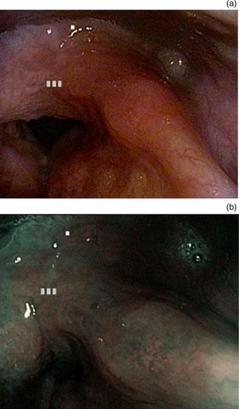
In all cases, laryngeal movements during laryngoscopy were normal, with no organic lesions observed under conventional white light or narrow-band imaging. The majority of patients (n = 31; 72 per cent), however, showed a certain degree of laryngeal hyperaemia, limited to the arytenoids in 23 cases (53.4 per cent) and more diffuse to the entire larynx in 8 cases (18.6 per cent). Thick endolaryngeal mucus was found in 10 patients (23.2 per cent) and laryngeal oedema in 17 patients (39.5 per cent; mild in 11 patients and moderate in 6 patients). Vocal fold oedema was generally slight and present in a minority of patients (n = 11; 25.6 per cent). Narrow-band imaging endoscopy confirmed the aforementioned features, showing particular enhancement of the microvessels and their dilation (Figure 1).
(a) Intense hyperaemia of the laryngeal vestibule, particularly in the arytenoids area, under conventional white light. (b) Identical anatomical image under narrow-band imaging; note the enhanced details of the microvessels of the left aryepiglottic fold, much more visible than during normal laryngoscopy.

The mean Reflux Finding Score was low (3.3 ± 3.2), with only seven patients (16.2 per cent) scoring 7 or higher. The Reflux Finding Score recalculated under narrow-band imaging corresponded with the conventional white light Reflux Finding Score.
Statistical analysis did not show any correlation between sex, age, smoking habit, disease duration, medication type, antineutrophil cytoplasmic antibodies status and the presence of laryngeal inflammation or a high Reflux Finding Score.
The literature review revealed only two articles on laryngeal involvement in eosinophilic granulomatosis with polyangiitis; further details are discussed below.
Discussion
Although a rare autoimmune disease, eosinophilic granulomatosis with polyangiitis may have a major impact on patients’ quality of life, because of systemic morbidity and organ-related involvement.Reference Groh, Pagnoux, Baldini, Bel, Bottero and Cottin1
Specialists should be aware of the expected morbidity in eosinophilic granulomatosis with polyangiitis patients and should explore all aspects of the disease. Laryngeal involvement in granulomatosis with polyangiitis is common. It is represented, in up to 15 per cent of patients, by hoarseness and stridor,Reference Rasmussen11, Reference Guerrero, Gall, Tilahun and Garon12 and is secondary, in all reported cases, to subglottic stenosis.Reference Belloso, Estrach and Keith5 Subglottic stenosis in granulomatosis with polyangiitis typically appears with dyspnoea when the stenosis involves more than 70 per cent of the airway; therefore, a pre-clinical subglottic stenosis diagnosis could be beneficial for patients and may prevent tracheostomy.Reference Gouveris, Karaiskaki, Koutsimpelas, Chongolwatana and Mann4
A low inflammatory status in the larynx is one of the goals of therapy in granulomatosis with polyangiitis cases. Indeed, ‘significant subglottic inflammation’ is one of the items checked by rheumatologists when determining the Birmingham Vasculitis Activity Score.Reference Luqmani, Bacon, Moots, Janssen, Pall and Emery8 Conversely, laryngeal involvement in eosinophilic granulomatosis with polyangiitis is not well documented.
The Birmingham Vasculitis Activity Score is also used in eosinophilic granulomatosis with polyangiitis patients to assess disease activity. We aimed to accurately explore the larynx, using the Birmingham Vasculitis Activity Score, to determine the prevalence of subglottic stenosis in a large cohort of eosinophilic granulomatosis with polyangiitis patients. Our study comprised one of the largest groups of patients reported in the literature, and the data were collected at a single institution in a European country. We demonstrated that laryngeal involvement in eosinophilic granulomatosis with polyangiitis can be considered uncommon or even exceptional; in fact, in over 43 patients, there were no cases of laryngeal paralysis or subglottic stenosis.
Our results are in line with current literature, which shows that even in large multicentre studies (n = 383 and n = 230),Reference Comarmond, Pagnoux, Khellaf, Cordier, Hamidou and Viallard13, Reference Guillevin, Pagnoux, Seror, Mahr, Mouthon and Le Toumelin14 no cases of laryngeal involvement were found. Indeed, Bacciu et al. assert that laryngeal involvement is ‘an exceedingly uncommon manifestation’ of eosinophilic granulomatosis with polyangiitis, with no cases reported in their cohort of 21 patients.Reference Bacciu, Bacciu, Mercante, Ingegnoli, Grasselli and Vaglio3
The literature review revealed only two case reports of eosinophilic granulomatosis with polyangiitis with laryngeal involvement. Mazzantini et al. reported persistent dysphonia with systemic symptoms (fever and arthralgia) in a 59-year-old patient, caused by right vocal fold paresis, with no visible lesions.Reference Mazzantini, Fattori, Matteucci, Gaeta and Ursino15 The authors hypothesised that the paresis could be secondary to peripheral neuropathy, but no histological demonstration of the presence of vasculitis was feasible. More recently, Al-Ammar et al. described the case of a nine-year-old girl who presented with a recurrent vegetating lesion involving the true vocal folds, with normal mobility and reduction of the respiratory space.Reference Al-Ammar, Yasin, Al-Muhsen, Al-Saadi and Al-Sohaibani16 The lesion was removed surgically and histology demonstrated the presence of a vasculitic process.
Our study provides interesting information on laryngeal inflammation in eosinophilic granulomatosis with polyangiitis patients. In our cohort, laryngeal damage was a minor entity and generally slight, with approximately 1 patient out of 10 showing significant laryngeal inflammation, more frequently arytenoid hyperaemia.
In our opinion, the Reflux Finding Score was very useful in providing a score for the clinical sign of ‘laryngeal inflammation’, enabling comparison among patients. In the literature, gastroesophageal involvement in eosinophilic granulomatosis with polyangiitis patients is a common feature: up to 50 per cent of these patients may have eosinophilic gastroenteritisReference Cojocaru, Cojocaru, Silosi and Vrabie17 or suffer from gastroesophageal reflux disease secondary to steroid therapy.Reference Pagnoux, Mahr, Cohen and Guillevin18 Unfortunately, in our group, the lack of data on the presence of gastroesophageal reflux disease makes it impossible to determine the relationship between Reflux Finding Score and gastroesophageal reflux disease.
We hypothesised a link between laryngeal inflammation and drug consumption, but statistical analysis did not reveal any significant associations between therapy, disease duration, anti-neutrophilic cytoplasmic antibodies status and the presence of laryngeal inflammation.
Narrow-band imaging is considered beneficial in evaluating laryngeal inflammation. Narrow-band imaging has a high sensitivity for detecting mucosal hyperaemia, possibly because of its ability to increase the contrast of blood vessels.Reference Cojocaru, Cojocaru, Silosi and Vrabie17 In our experience, narrow-band imaging and conventional white light showed the same details, with no differences in terms of Reflux Finding Score. In our opinion, the narrow-band imaging related advantage was not detectable because of the small sample size and because of the large majority of normal larynxes included in the study.
Our experience shows that laryngeal inflammation is a common feature among eosinophilic granulomatosis with polyangiitis patients. However, only a minority of the patients had severe inflammation, as per the Reflux Finding Score (determined by conventional white light and narrow-band imaging). Primary laryngeal involvement in eosinophilic granulomatosis with polyangiitis appears to be very uncommon, as evidenced by our group and the literature review. This suggests that the importance of laryngeal involvement in assessing disease activity for the Birmingham Vasculitis Activity Score should be reconsidered. All specialists involved in eosinophilic granulomatosis with polyangiitis management should keep our experience in mind.
• Eosinophilic granulomatosis with polyangiitis is a systemic necrotising vasculitis
• It is associated with ENT involvement, especially in the nose and paranasal sinuses
• Less is known about laryngeal involvement: a literature review revealed two eosinophilic granulomatosis with polyangiitis cases with laryngeal involvement
• Laryngeal inflammation appears to be a common feature, by means of laryngitis or gastroesophageal reflux
• Primary laryngeal involvement in eosinophilic granulomatosis with polyangiitis seems very uncommon
• Its importance in assessing disease activity in the Birmingham Vasculitis Activity Score should be reconsidered
Acknowledgements
We thank Mrs Diana Elizabeth Hearn for her support in revising and translating the manuscript, and Ms Anna Giovinazzo for her great help in organising the scientific work.
Competing interests
None declared.