Anomalous origin of the left coronary artery from the main pulmonary artery in univentricular hearts is a rare congenital anomaly. It is especially rare for the coronary artery to arise from the right pulmonary artery. The combination of these anomalies has been described in the literature; however, the prevalence of such a condition is unknown.Reference Turiy, Douglas and Balaguru 1 – Reference Malec, Mroczek, Pajak, Zajac, Kołcz and Januszewska 6 Being able to recognise it and deal with it intraoperatively is very challenging. We report the case of a neonate with a univentricular heart in the form of a double-outlet right ventricle, mitral atresia with discrete coarctation of the aorta, and an incidental intraoperative finding of an anomalous origin of the left coronary artery from the right pulmonary artery who underwent a successful surgical repair.
Case report
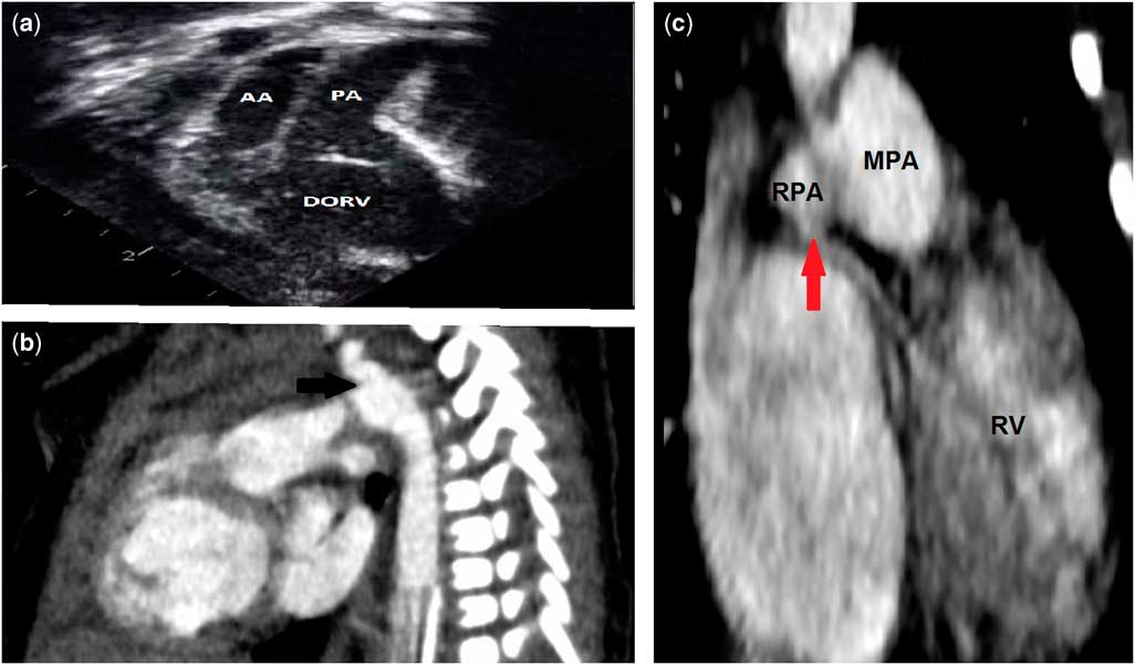
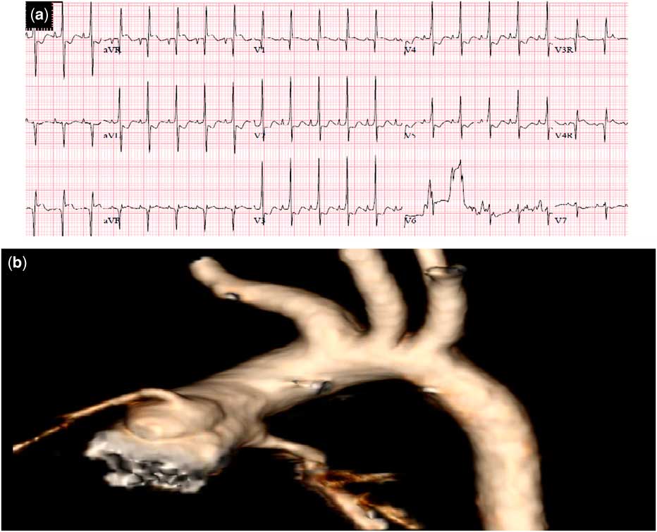
A baby girl was born at 39 weeks (3 kg) with a prenatal diagnosis of a double-outlet right ventricle, mitral atresia, and a functionally univentricular heart with possible coarctation of the aorta (Fig 1a). The baby was started on prostaglandins and transferred to the cardiovascular ICU. Upon arrival at the unit, the patient was found to be comfortable on room air with oxygen saturations in the low- to mid-80s. A postnatal echocardiography was performed, which revealed mitral-valve atresia, a double-outlet right ventricle with side-by-side great arteries, one coronary artery visualised as arising from the anterior and leftward coronary sinus, and the aortic arch and isthmus within the normal measurement for body surface area with a large patent ductus arteriosus. Prostaglandins were stopped on day-of-life 2. A CT scan was performed on day-of-life 3 to assess the coronary arteries but unfortunately they could not be inspected despite the retrospective technique and extensive motion correction for the patient’s rapid heart rate. The aortic isthmus was mildly hypoplastic, measuring 3.9×5.0 mm proximal to the insertion of a small patent ductus arteriosus (Fig 1b). Of note, a small vessel coursing leftwards along the under-surface of the proximal right pulmonary artery was interpreted as a venous structure. This vessel was found intraoperatively to be an anomalous left coronary artery arising from the right pulmonary artery (Fig 1c). A period of 48 hours after stopping the prostaglandins, the patient showed clinical evidence of decompensated heart failure and ST segment elevation with tachycardia thought to be related to pulmonary over-circulation (Fig 2a). Surprisingly, she also developed progressive tricuspid regurgitation and ventricular dysfunction. She was taken to the operating room urgently for pulmonary artery banding and coarctation repair.
( a ) Preoperative echocardiogram showing double-outlet right ventricle (DORV) with side-by-side great arteries and mitral atresia (univentricular heart). ( b ) Preoperative CT scan showing the narrowed aortic isthmus (black arrow). ( c ) Preoperative CT scan showing a small vessel (red arrow) beneath the right pulmonary artery. In retrospect, this vessel is the anomalous left coronary artery arising from the right pulmonary artery. AA=ascending aorta; MPA=main pulmonary artery; PA=pulmonary artery; RPA=right pulmonary artery; RV=right ventricle.

( a ) A preoperative electrocardiogram showing ST elevation in the inferior leads and ST depression in the anterolateral leads. ( b ) A postoperative CT scan with three-dimensional reconstruction showing the two coronary systems with the reconstructed aorta.

In the operating room, a median sternotomy was performed and we noted significant irritability of the ventricle. The pericardium was opened and the heart was noted to be severely dilated with relatively poor function. The ST segments were profoundly abnormal during that time. We knew from the preoperative echocardiogram and CT scan that the duct was small. We quickly placed a loose pulmonary artery band on the main pulmonary artery with the hope of improving the haemodynamics. The systemic blood pressure rose initially, suggesting that there was some pulmonary over-circulation, but in very short order the haemodynamics deteriorated. The ductus arteriosus was temporarily snared without any change in the haemodynamics. We felt that perhaps we could band the branched pulmonary arteries more accurately than we could the main pulmonary artery and we began to mobilise the branched pulmonary arteries. This is when we identified the anomalous left coronary artery arising from the right pulmonary artery. Once the branched pulmonary arteries were banded distal to the coronary artery, we noted a dramatic improvement in the haemodynamics, myocardial perfusion, and near resolution of the ST segment abnormalities. At this point, we considered leaving the mild coarctation alone and placing permanent bilateral pulmonary artery bands, but we were concerned that this would be difficult to achieve. The decision was made to repair the coarctation and transfer the left coronary artery to the aortic root. After heparinisation, a 3.5 mm Gore-Tex graft was sewn to the side of the distal innominate artery. The graft was cannulated, secured, de-aired and connected to the arterial limb of the bypass circuit. The right atrium was cannulated directly, and cardiopulmonary bypass was initiated. The temperature was reduced to 18°C and further mobilisation of the branched pulmonary arteries and the anomalous coronary artery was undertaken. The ascending aorta was cross-clamped as distally as possible, and cold crystalloid cardioplegia was administered anterograde through the aortic root. A partial arrest followed and the ductus arteriosus was ligated. The right pulmonary artery was opened on its anterior aspect and cold crystalloid cardioplegia was administered directly into the anomalous left coronary artery. The left coronary artery was mobilised from the right pulmonary artery on a large button. The ascending aorta was opened longitudinally on its anterior aspect and a 4 mm defect was created posteriorly at the sinotubular junction with an aortic punch. The anomalous left coronary artery was anastomosed to the aorta and the incision on the anterior wall of the aorta was then closed primarily. Cardioplegia was re-administered, demonstrating patency of the coronary valve and competency of the aortic valve. Under a period of selective cerebral perfusion, the isthmus was transected and the minor curvature of the distal arch was incised longitudinally. The descending aorta was extensively mobilised and all ductal tissue was removed. An extended end-to-end anastomosis was then performed. The right pulmonary artery defect was repaired with a fresh autologous pericardial patch. After re-warming, the heart was de-aired and the aortic cross-clamp was removed. A normal sinus rhythm had returned rapidly and we completed the re-warming process. A pulmonary artery band was placed on the main pulmonary artery and secured to prevent distal migration. The patient was weaned from bypass and put on moderate inotropic support. We noted excellent haemodynamics. The baby did well postoperatively and was discharged home on postoperative day 31. A pre-Glenn CT scan was performed, which showed patent coronary arteries and the aorta (Fig 2b).
Comment
Anomalous origin of the left coronary artery from the right pulmonary artery in univentricular hearts is a very rare congenital anomaly. Few studies have described this anomaly in the literature. Most cases were associated with hypoplastic left heart syndrome.Reference Sarris, Drummond-Webb, Ebeid, Latson and Mee 2 , Reference Nosal, Omeje and Poruban 4 – Reference Malec, Mroczek, Pajak, Zajac, Kołcz and Januszewska 6 One study described a double-outlet right ventricle with mitral atresia and a left circumflex artery from the right pulmonary artery.Reference Nathan, Emani, Marx and Pigula 3 Other case reports described the anomalous artery to be originating from the main pulmonary artery rather than from its branches.Reference Turiy, Douglas and Balaguru 1 , Reference Nathan, Emani, Marx and Pigula 3 It has also been described in biventricular hearts.Reference Saroli, Gelehrter, Gomez-Fifer, van der Velde, Bove and Ensing 7 , Reference Bitar, Kveselis, Smith, Byrum and Quaegebeur 8
It can be diagnosed preoperatively using echocardiography, cardiac catheterisation, or CT angiography.Reference Sarris, Drummond-Webb, Ebeid, Latson and Mee 2 , Reference Nathan, Emani, Marx and Pigula 3 , Reference Saroli, Gelehrter, Gomez-Fifer, van der Velde, Bove and Ensing 7 When it is discovered preoperatively, the outcome is much better with prolonged survival.Reference Sarris, Drummond-Webb, Ebeid, Latson and Mee 2 , Reference Nathan, Emani, Marx and Pigula 3 , Reference Saroli, Gelehrter, Gomez-Fifer, van der Velde, Bove and Ensing 7 , Reference Bitar, Kveselis, Smith, Byrum and Quaegebeur 8 Failure to diagnose it before surgery may complicate the intraoperative and postoperative courses. It has been shown that the intraoperative recognition of this anomaly after instituting cardiopulmonary bypass will most likely lead to adverse outcomes and eventually death.Reference Turiy, Douglas and Balaguru 1 , Reference Nathan, Emani, Marx and Pigula 3 , Reference Jaworski, Kansy and Laniewski-Wollk 5 , Reference Malec, Mroczek, Pajak, Zajac, Kołcz and Januszewska 6 Only one case has been reported in the literature in which the patient survived after bypass initiation and intraoperative recognition.Reference Nosal, Omeje and Poruban 4
Unfortunately, the preoperative CT angiogram for this patient was misinterpreted by both the radiologist and the surgeon and the correct diagnosis was missed. At our centre, we utilise a retrospective electrocardiogram-gated CT scan to delineate the coronary anatomy. A three-dimensional workstation is used for post-processing analysis, including the generation of maximum intensity projection images. To obtain clear images of the coronary arteries, the patient’s heart rate should be well controlled. This was not the case in our patient as she had a very fast heart rate, and post-processing analysis could not overcome the problem.
Despite being a very rare lesion, failure to recognise it may lead to disastrous outcomes, including death. Factors that may help in its recognition intraoperatively include absence of a left coronary system arising from the aorta upon inspection, failure to attain complete diastolic cardiac arrest when administering cardioplegia, discolouration of the myocardium, and poor ventricular function when being taken off bypass. This case shows the importance of being familiar with the coronary anatomy in single-ventricle hearts.
The intraoperative discovery of this anomaly was surprising and necessitated a complete understanding of the disease physiology that caused the haemodynamic collapse upon applying the pulmonary artery band. Applying the pulmonary artery band initially was potentially unsafe and underscores the importance of correct diagnosis in preoperative planning. Knowing that the patient had ST segment changes preoperatively, with inability to visualise the coronaries on the CT scan, the safest approach would have been imaging the coronaries in the catheterisation lab before surgery.
To our knowledge, this the first report of a neonate with a univentricular heart in the form of a double-outlet right ventricle, mitral atresia with discrete coarctation of the aorta, and intraoperative finding of an anomalous origin of the left coronary artery from the right pulmonary artery who underwent a successful repair and had an uneventful postoperative course.
Acknowledgements
The authors would like to thank Dr Luis De Leon for providing the CT angiography image used in this paper. The parents granted permission to publish the case report. Authors’ contributions: All authors participated in creating content for the manuscript as well as in editing, and provided final approval for submission.
Financial Support
This research received no specific grant from any funding agency or from any commercial or not-for-profit sectors.
Conflicts of Interest
The authors declare no potential conflicts of interest with respect to the study, authorship, and/or publication of this article.