Introduction
Malaria is caused by the Plasmodium falciparum parasite, which is transmitted from person to person by the bite of an infected anopheles mosquito.Reference Batey, Lightbown, Lambert, Edwards and Coker1 There are four types of malaria; of these, two are more common, with symptoms including fever, chills, headache, muscle aches, sweating and shivering, and malaise, and symptoms of progressive anaemia.Reference Baune, Flinois, Furlan, Taburet, Becquemont and Farlnotti2
Halofantrine is a newly developed antimalarial drug proposed to replace the older quinine group drugs.Reference Ter Kuile, Dolan, Nosten, Edstein, Luxemburger and Phaipun3, Reference White4 It is effective in the treatment of acute malaria caused by single or mixed infections of P falciparum or P vivax.Reference Baune, Flinois, Furlan, Taburet, Becquemont and Farlnotti2 It can also be used to treat mild to moderately severe chloroquine-resistant and multidrug-resistant malarial infections.Reference Connell and Emilio5, Reference Nosten, McGready, d'Aleessandro, Nonell, Verhoeff and Menenez6
The introduction of this drug has been delayed because of its possible side effects, with insufficient studies on adverse reactions in humans.Reference Ter Kuile, Dolan, Nosten, Edstein, Luxemburger and Phaipun3
Halofantrine acts by forming toxic complexes with ferritoporphyrin IX which damage the parasite membrane. Peak plasma concentrations are achieved 4–8 hours after oral intake.Reference Nosten, McGready, d'Aleessandro, Nonell, Verhoeff and Menenez6, Reference Nyunt and Plowe7 Halofantrine is generally well tolerated, with a low incidence of gastrointestinal side effects.Reference Ter Kuile, Dolan, Nosten, Edstein, Luxemburger and Phaipun3
Halofantrine is administered as an 8 mg/kg treatment divided into three doses and repeated after 7 days.Reference Nosten, McGready, d'Aleessandro, Nonell, Verhoeff and Menenez6
The known ototoxic effects of antimalarial drugs include: vertigo; tinnitus; and a transient, reversible, bilateral, symmetrical, sensorineural hearing loss with a characteristic audiographic notch at 4 kHz.Reference Jung and Rhee8, Reference Winstanley and Ward9 These side effects are probably secondary to decreased perfusion to and biochemical alterations in the cochlea, together with direct damage to the outer hair cells.Reference Miller10, Reference Makanga, Premji, Falade, Karbwang, Mueller and Andriano11 Most antimalarial drugs show marked, reversible ototoxic effects, but halofantrine appears to be less ototoxic than quinine.Reference Nosten, McGready, d'Aleessandro, Nonell, Verhoeff and Menenez6, Reference Nyunt and Plowe7
To our knowledge, however, no previous studies have investigated the effect of halofantrine on hearing. Thus, we conducted the present study to evaluate the effects of halofantrine on the cochlea of guinea pigs, using different histological preparations.
Materials and methods
This study was conducted in the ENT and histological units of Suez Canal University Hospital, Ismailia, Egypt. Healthy pigmented guinea pigs (male or female) of the same age and average weight (approximately 200 g) were used, selected according to the following criteria: a normal eardrum (as per otoscopic examination, to exclude infection) and a preserved pinna reflex (i.e. reflex contraction of the auricle in response to loud sounds, a rough indication of normal hearing).
Thirty guinea pigs were divided into three groups of 10: a control group, a halofantrine therapeutic dose group, and a halofantrine double therapeutic dose group.
The 10 control group guinea pigs received 0.05 ml of physiological saline/100 g body weight, divided into three doses given orally at 6 hourly intervals.
The 10 guinea pigs in the halofantrine therapeutic dose group received 0.05 ml of halofantrine/100 g body weight, which corresponded to a therapeutic dose of 8 mg/kg body weight, divided into three doses given orally at 6 hourly intervals.Reference Nosten, McGready, d'Aleessandro, Nonell, Verhoeff and Menenez6
The 10 guinea pigs in the halofantrine double therapeutic dose group received 0.10 ml of halofantrine/100 g body weight, which corresponded to twice the therapeutic dose of 8 mg/kg body weight, divided into three doses given orally at 6 hourly intervals.
Drugs were administered orally dissolved in saline, 2 hours after food (as dosing with food, especially fatty food, has been shown to increase the rate of absorption, with an increased risk of drug side effects such as irregular heart rate).Reference Nosten, McGready, d'Aleessandro, Nonell, Verhoeff and Menenez6
The guinea pigs were sacrificed 12 hours after the last drug dose. All animals received an intraperitoneal injection of 100 mg sodium pentobarbital, followed if necessary by a 50 mg intracardiac injection after loss of consciousness.
Approval for similar experimental studies had previously been obtained from the animal care and use committee of our institution.Reference Yung and Wright12
The animals' temporal bones were placed in ethylene diamine triacetic acid solution for 25 days. Specimens were processed into 6 mm thick sections.
Histological techniques
One cochlea from each animal was stained with haematoxylin and eosin (H&E) and the other with toluidine blue.
In order to visualise the general architecture of the inner ear, Harris's haematoxylin was prepared with the following: 2.5 g haematoxylin, 2.5 ml absolute ethyl alcohol, 50 g potassium alum, 500 ml distilled water, 1.25 g mercuric oxide and 20 ml glacial acetic acid.
Haematoxylin was dissolved in absolute alcohol and added to the alum, which had previously been dissolved in warm distilled water. The mixture was rapidly brought to the boil and mercuric oxide added. The solution was rapidly cooled by plunging the flask into cold water. When the solution was cold, acetic acid was added, and the stain was ready for immediate use. Glacial acetic acid was used to give more precise, selective staining of nuclei.
Eosin was prepared as a 1 per cent solution in distilled water.
The H&E staining technique proceeded as follows: (1) dewaxing of paraffin sections in xylol and then hydration through graded alcohol–water mixtures; (2) haematoxylin staining for 5 minutes; (3) washing under running tap water until specimens became bluish; (4) differentiation in 1 per cent acid alcohol (5–10 seconds); (5) washing with tap water until bluish again; (6) eosin counter-staining for 2 minutes; (7) rapid rinsing; and (8) dehydration with ascending grades of alcohol, clearing in xylene and mounting in distyrene-plasticiser-xylene mixture.Reference Steven, Bancroft and Stevens13
Toluidine blue stain was prepared with 0.2 per cent aqueous toluidine blue and 95 per cent ethyl alcohol.
This staining technique proceeded as follows: (1) dewaxing of paraffin sections in xylene and progressive hydration down to distilled water (through graded alcohol–water mixtures); (2) toluidine blue staining for 6 hours at room temperature; (3) washing in distilled water; (4) rinsing in 90 per cent alcohol and differentiation in 95 per cent alcohol, controlled microscopically until Nissl's substances were seen in the cell bodies of large neurons; and (5) dehydration in alcohol, clearing in xylene and mounting in distyrene-plasticiser-xylene mixture.Reference Bancroft, Cook, Sterling and Turner14
Statistical analysis
Histological data were collected using both staining techniques for all three animals groups, and were then analysed and compared with normal images.
Results and analysis
The guinea pig cochlea is normally greyish-yellow in colour and approximately 3 × 4 mm in size. It has a wide base and tapering apex. It has the appearance of a spiral formed of three and a half turns encircling a bony core (the modiolus) visible on longitudinal sections.
The guinea pig cochlear duct appears triangular; its roof consists of a thin membrane (Reissner's membrane), while its floor is composed of an outer bony spiral lamina from the modiolus and an inner basilar membrane, which carries the organ of Corti on its upper surface.
Control group
No gross changes were detected in control group cochlear specimens.
Haematoxylin and eosin
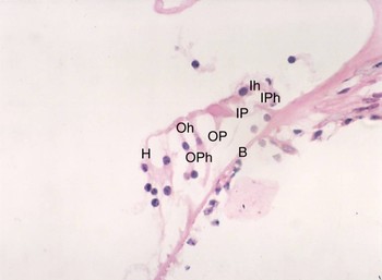
In specimens treated with this stain, the cochlea appeared as a cone-like structure with a pointed apex and wide base. Each turn was divided into three components, the scala vestibuli, scala media and scala tympani, as seen in Figure 1. The normal structures appeared as follows: nuclei stained blue; cytoplasm stained varying shades of pink; muscle fibres stained a deep pink-red; and collagen stained a pale pink-red. The three rows of outer hair cells appeared as tall cells with acidophilic cytoplasm and basal, basophilic nuclei. One row of the inner hair cells showed acidophilic cytoplasm but more globular cell bodies, as seen in Figure 2.
Photomicrograph of cochlea of a control group guinea pig, showing the structure of the organ of Corti. B = basilar membrane; O = organ of Corti; S = stria vascularis; L = limbus; T = tectorial membrane; V = vestibular or Reissner's membrane; SG = spiral ganglion; SL = spiral ligament; SV = scala vestibule; SM = scala media; ST = scala tympani. (H&E; ×100)

Photomicrograph of cochlea of a control group guinea pig, showing the detailed structure of the organ of Corti. IP = inner pillar cell; IPh = inner phalangeal cell; Ih = inner hair cell; OP = outer pillar cell; OPh = outer phalangeal cell; Oh = outer hair cell; H = Hensen's cell; B = basilar membrane. (H&E; ×400)

Toluidine blue
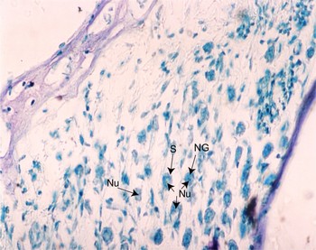
Spiral ganglia sections from control group specimens treated with this stain revealed aggregates of dark blue granules (Nissl's granules), together with pale-staining rounded areas (representing nuclei) containing deep blue stained nucleoli, as seen in Figure 3.
Photomicrograph of cochlea of a control group guinea pig, showing spiral ganglion cells (S), nuclei (Nu) and Nissl's granules (NG) filling the cytoplasm. (Toluidine blue; ×400)

Halofantrine therapeutic dose group
This group received 0.05 ml of halofantrine/100 g body weight, which corresponded to the therapeutic dose of 8 mg/kg body weight, divided into three doses and given orally at 6 hourly intervals.Reference Nosten, McGready, d'Aleessandro, Nonell, Verhoeff and Menenez6
Haematoxylin and eosin
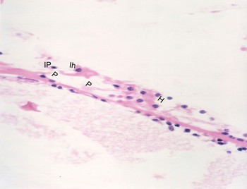
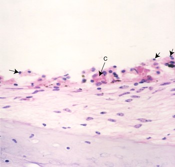
Specimens stained with H&E showed loss and distortion of inner hair cells and inner phalangeal cells, rupture and distortion of pillar cells, and rupture and vacuolation of Hensen's cells, as seen in Figure 4. These specimens also showed atrophy of the stria vascularis, observed as cell loss and distorted cellularity, together with separation of the stria vascularis from the underlying spiral ligament, and congestion and fusion of stria vascularis blood vessels, as seen in Figure 5.
Photomicrograph of cochlea of a guinea pig in the halofantrine therapeutic dose group, showing rupture of Hensen's cells (H) and pillar cells (P), in addition to loss of inner phalangeal cells (IP) and inner hair cells (Ih). (H&E; ×400)

Photomicrograph of cochlea of a guinea pig in the halofantrine therapeutic dose group, showing congestion and fusion of blood vessels in the stria vascularis (C), distorted cellularity, and loss of cells (→), in addition to separation from the underlying spiral ligament (∧ ∧ ∧). (H&E; ×400)

Toluidine blue
Toluidine blue stained spiral ganglia sections showed chromatolysis, giving the cytoplasm a pale appearance due to reduced Nissl's granules, with moderate loss of spiral ganglia cells, as seen in Figure 6.
Photomicrograph of cochlea of a guinea pig in the halofantrine therapeutic dose group, showing mild loss of spiral ganglion cells in addition to chromatolysis (loss of Nissl's granules) in some cells (→). (Toluidine blue; ×400)

Halofantrine double therapeutic dose group
Ten guinea pigs received 0.10 ml of halofantrine/100 g body weight, which corresponded to twice the therapeutic dose of 8 mg/kg body weight, divided into three doses given orally at 6 hourly intervals.
Haematoxylin and eosin
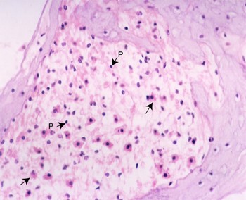
This staining shows distortion and loss in inner and outer hair cells and inner and outer phalangeal cells. Pillar cells showed rupture and distortion, with vacuolation of Hensen's cells. Congestion of blood vessels was observed, together with atrophy of the stria vascularis in the form of distorted cellularity, cell loss and pyknosis, as seen in Figure 7. Spiral ganglia sections showed a decrease in cell numbers and light staining of spiral ganglion cell cytoplasm, as seen in Figure 8.
Photomicrograph of cochlea of a guinea pig in the halofantrine double therapeutic dose group, showing congestion of blood vessels (C) and distorted cellularity with pyknotic nuclei (→) and cell loss. (H&E; ×400)

Photomicrograph of cochlea of a guinea pig in the halofantrine double therapeutic dose group, showing moderate to severe loss of spiral ganglion cells, reduced staining (→) and pyknotic nuclei (P). (H&E; ×400)

Toluidine blue stain
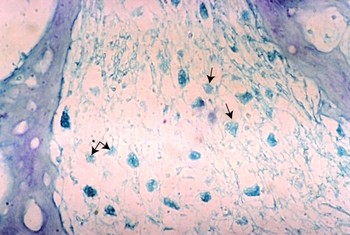
Spiral ganglia sections stained with toluidine blue showed chromatolysis, giving the cytoplasm a pale appearance due to reduced number of Nissl's granules, and severe loss of spiral ganglia cells, as seen in Figure 9.
Photomicrograph of cochlea of a guinea pig in the halofantrine double therapeutic dose group, showing severe loss of spiral ganglion cells in addition to chromatolysis (with loss of Nissl's granules) in some cells (→). (Toluidine blue; ×400)

Discussion
Halofantrine is a phenanthrene methanol derivative related to mefloquine and quinine.Reference Connell and Emilio5 This newly developed antimalarial drug is proposed as a replacement for the older quinine group drugs, due to increasing reports of side effects from the latter drugs.Reference Ter Kuile, Dolan, Nosten, Edstein, Luxemburger and Phaipun3 The US Food and Drug Administration approved halofantrine for use in 1992.Reference Touze, Fourcade, Peyrone, Heno and Deharo15
The introduction of halofantrine has been delayed because of fears of side effects and insufficient studies on adverse reactions in humans.Reference Ter Kuile, Dolan, Nosten, Edstein, Luxemburger and Phaipun3 Older antimalarial drugs have been shown to have marked, reversible ototoxic effects.Reference Horton16 However, there are currently no studies investigating the effects of halofantrine on the cochlea. However, the clinical experience of the otolaryngology staff of Suez Canal University within endemic areas such as the Republic of Yemen (where malaria is a more common health problem) has indicated that the drug may cause tinnitus and transient hearing loss.
Different doses of halofantrine were used in our study. The recommended therapeutic dose of 8 mg/kg/day was used in the halofantrine therapeutic dose group to investigate cochlear histopathological changes, while double the therapeutic dosage was used in the third animal group to demonstrate dose-dependent effects of halofantrine on cochlear structures. Both doses were adjusted according to the animal's body weight.Reference Touze, Fourcade, Peyrone, Heno and Deharo15
The halofantrine therapeutic dose group showed inner hair cell loss with distortion, inner phalangeal cell loss, rupture of pillar cells and Hensen's cells, and decreased numbers of spiral ganglia cells. In their study of the effect of halofantrine on rat testes, Diadia et al. found a reduction in the number and level of maturity of spermatogenic cells, associated with damage to the seminiferous tubules, following animal exposure to therapeutic doses of halofantrine.Reference Diadia, Gbigbi and Dapper17
In addition, we observed degeneration of the spiral ganglia cells, in the form of chromatolysis. Halofantrine has also been shown to have hepatotoxic effects; Obi et al. found pathological hepatic degeneration following exposure of guinea pigs to halofantrine.Reference Obi, Orisakwe, Asomugha, Udemezue and Orish18
The halofantrine double therapeutic dose group showed atrophy of the stria vascularis, in the form of cell loss, pyknosis and distorted cellularity. The stria vascularis also showed congestion of blood vessels. These findings are in accordance with those of Obi et al., who found portal triaditis (i.e. inflammation of the portal tract triad including the portal vein, hepatic artery and bile duct) following exposure of guinea pigs to halofantrine.Reference Obi, Orisakwe, Asomugha, Udemezue and Orish18
In the halofantrine double therapeutic dose group, we also noted inner and outer hair cell distortion and loss, inner and outer phalangeal cell distortion and loss, rupture and distortion of pillar cells, and rupture of Hensen's cells. These severe effects, with massive cell loss, are in accordance with Diadia and colleagues' findings in spermatogenic cells: following exposure to halofantrine at double the therapeutic dose, these authors found disturbed seminiferous tubule cellularity, in the form of cell number reduction, loss of maturity and degeneration.Reference Diadia, Gbigbi and Dapper17
Our findings of outer hair cell distortion and loss are similar to those of Berninger and Gustafsson, who observed damage to the outer hair cells of the organ of Corti following exposure to quinine.Reference Berninger and Gustafsson19 Halofantrine and quinine share many side effects, including neurological, vestibular, visual and abdominal problems.Reference Touze, Fourcade, Peyrone, Heno and Deharo15 The correlation between the observed histopathological effects of halofantrine and quinine on outer hair cells may be due to their structural similarity.Reference Berninger and Gustafsson19
• Halofantrine is a new antimalarial drug proposed to replace the older quinine group of drugs
• Introduction of this drug has been delayed due to fears of its side effects and insufficient studies on adverse reactions in humans
• This study demonstrated histological signs of ototoxicity following halofantrine exposure, in a guinea pig cochlea model
In our sections, the stria vascularis showed congestion of blood vessels and atrophy, in the form of distorted cellularity, cell loss and pyknosis. These features could be explained by the findings of Diadia et al., who reported tubular atrophy and degeneration of seminiferous tubules following exposure to double-dose halofantrine in an animal model.Reference Diadia, Gbigbi and Dapper17
In our histological preparations, spiral ganglia sections showed decreased cell numbers and cell degeneration in the form of chromatolysis.
In addition, all of our above-mentioned findings are in agreement with those of Bassi et al., who found that halofantrine exhibited cardiotoxic and hepatotoxic effects.Reference Bassi, Buratai and Kuchali20
Based on the above data, it was impossible to determine whether the ototoxic effects of halofantrine were reversible or not.
Conclusion
In a guinea pig model, the recommended therapeutic dose of halofantrine results in mild to moderate histological changes in the cochlea; at double the therapeutic dose, marked histological changes occur. Thus, halofantrine may be considered an ototoxic drug.