The reproduction, variation and spread of harmful microorganisms have severe impacts on human health and environmental sanitation, as exemplified by outbreaks of severe acute respiratory syndrome (SARS) and novel coronavirus pneumonia (COVID-19), both caused by coronavirus variants (Imani et al., Reference Imani, Ladouceur, Marshall, Maclachlan, Soleymani and Didar2020; Nguyen et al., Reference Nguyen, Nguyen, Nguyen, Nguyen, Nguyen and Tran2022). In addition, infections induced by pathogenic bacteria, including Escherichia coli, Staphylococcus aureus, Pseudomonas aeruginosa, Klebsiella pneumoniae and Streptococcus pyogenes, as well as various fungi, continue to present significant threats to global public health (Garmendia et al., Reference Garmendia, Frankel and Crepin2005; Ye et al., Reference Ye, Li, Li, Wang, Mao and Xie2011; Song et al., Reference Song, Tang, Ding, Tan, Zhang and Wang2024). Therefore, various antimicrobial materials have been developed and applied in the field of human health and environmental safety to effectively inhibit the growth and reproduction of harmful microorganisms and reduce the spread of infectious diseases. Among these, inorganic antimicrobial materials have attracted significant attention due to their high efficiency, broad-spectrum antimicrobial activity, environmental friendliness and low potential for inducing drug resistance (Hoseinnejad et al., Reference Hoseinnejad, Jafari and Katouzian2018; Shaw et al., Reference Shaw, Kuriakose, Cheeseman, Dickey, Genzer and Christofferson2021). In particular, magnesium oxide (MgO) nanocrystals have attracted especial interest due to their excellent stability, prolonged antimicrobial efficacy and low cytotoxicity to biological cells, making them promising candidates for antimicrobial materials (Feng et al., Reference Feng, Wang, Jin, Zhang, Huang and Gao2016; Nguyen et al., Reference Nguyen, Grelling, Wetteland, Rosario and Liu2018). Rajagopalachar et al. (Reference Rajagopalachar, Pattar and Mulla2022) synthesized MgO nanosheets with a high specific surface area via a co-precipitation method. The MgO nanosheets exhibited significant antibacterial activity against both Bacillus cereus and P. aeruginosa. Krishnamoorthy et al. (Reference Krishnamoorthy, Manivannan, Kim, Jeyasubramanian and Premanathan2012) observed that the antibacterial activity of nano-MgO particles is closely related to the number of oxygen vacancy defects on the surface. As the particle size of nano-MgO decreased, the number of oxygen vacancy defects gradually increased, leading to a higher concentration of generated ·O₂⁻ and a consequent significant improvement in antimicrobial activity (Leung et al., Reference Leung, Ng, Xu, Shen, Gethings and Wong2014). However, such smaller particle sizes also result in more pronounced agglomeration, which limits the antimicrobial efficacy of MgO nanoparticles (NPs). To reduce this agglomeration, Arora et al. (Reference Arora, Sidhu and Bala2021) utilized the large specific surface area of sepiolite to achieve the uniform dispersion of MgO particles on its surface. This method not only achieved a uniform distribution of magnesium oxide NPs, but also exposed a greater number of active sites, thereby increasing the ability of MgO to inhibit spore germination and disrupt fungal structures, which effectively reduced the risk of fungal infection in rice plants. In addition, Esteban-Cubillo et al. (Reference Esteban-Cubillo, Pecharromán, Aguilar, Santarén and Moya2006) reported an antimicrobial nanocomposite formed by embedding Cu NPs into sepiolite mineral, which reduced S. aureus and E. coli concentrations by 99.9%. The synthesis process was the exchange of Cu ions with a proportion of the Mg ions located at the octahedral sites of sepiolite, followed by the formation of monodispersed Cu NPs decorated on the sepiolite surface. Therefore, these findings demonstrated that sepiolite minerals are attractive carrier materials owing to their fibre nanomorphology, large specific surface area, abundant pore structure and low biotoxicity (Zhang et al., Reference Zhang, Han, Zhao, Li, Meng, Zhang and Liang2022).
Sepiolite is a naturally occurring hydrated magnesium silicate characterized by high silica and magnesium contents as well as a unique fibrous structure. Based on these features, Li et al. (Reference Li, Nie, Tang, Tian and Liang2022a) utilized sepiolite as a template for the in situ synthesis of copper silicate composites with a duster-like hollow mesoporous structure. Similarly, Han et al. (Reference Han, Zhang, Zhang, Zhao, Zhang and Liang2021) prepared nano-zero-valent iron/sepiolite composite materials using a facile and environmentally friendly method. In this study, sepiolite as a supporter effectively improved the dispersion of nano-zero-valent iron particles and increased the contact area between contaminants and active sites, resulting in improved catalytic performance than pure nano-zero-valent iron. Jiang et al. (Reference Jiang, Peng, Yang, Li, Tang and Y.H2024) further fabricated a series of sepiolite-based nanocomposites with enhanced antibacterial properties, including sepiolite-Ag (Sep/Ag), sepiolite-CuO (Sep/CuO) and sepiolite-ZnO (Sep/ZnO). Among these, the Sep/Ag composite exhibited 100% antibacterial efficacy at a low Ag content of only 5%. These studies demonstrate that the high magnesium content and the unique fibrous structure of sepiolite enable it to serve both as a magnesium source and as a functional carrier material. In addition to its function as a carrier material, the sepiolite mineral possesses natural antimicrobial properties, attributed to its excellent adsorption performance, favourable exchangeable cations and surface electrical behaviour (Williams et al., Reference Williams, Metge, Eberl, Harvey, Turner, Prapaipong and Poret-Peterson2011; Ben Haj Yahia et al., Reference Ben Haj Yahia, Labille and Schneider2025). These characteristics make sepiolite a promising candidate for the synthesis of MgO/sepiolite composite antimicrobial materials. However, to the best of our knowledge, the preparation of nano-MgO/sepiolite composites from sepiolite without the addition of external magnesium salts has not been reported previously.
In this study, nano-MgO/sepiolite composite materials were prepared via an acid leaching–hydrothermal combined process using sepiolite as both the magnesium source and carrier material, and their antimicrobial activity against bacteria and fungi was evaluated. The main objectives were to: (1) develop a novel synthesis method for nano-MgO/sepiolite composite materials based on the crystal structure and chemical composition characteristics of sepiolite; (2) evaluate the antimicrobial performance of nano-MgO/sepiolite composite materials; and (3) investigate the underlying antimicrobial mechanism of the nano-MgO/sepiolite composite materials against bacteria and fungi. This study presents a novel strategy for developing highly effective inorganic antimicrobial agents through the high-value utilization of natural mineral materials. The developed composite materials exhibit considerable advantages for antimicrobial applications, particularly for creating healthy indoor environments.
Experimental
Materials and methods
The sepiolite clay mineral used in this experiment was form Xiangtan (Hunan Province, China). Hydrochloric acid (HCl; 36 wt.%) and sodium hydroxide (NaOH; analytical reagent (AR)) were purchased from Kelmel Chemical Reagents Co., Ltd. Saline solution was provided by Shijiazhuang No. 4 Pharmaceutical Co., Ltd. Agar powder and broth culture medium were supplied by Beijing Aoboxing Biotechnology Co., Ltd. The strains used in this experiment were E. coli and S. aureus, purchased from Beijing Baocang Biotechnology Co., Ltd. Deionized water was used throughout the experiment.
Synthesis of nano-MgO/sepiolite composites
Sepiolite was treated via an acid leaching–hydrothermal combined method to prepare nano-MgO/sepiolite composites. Firstly, diluted HCl solutions with concentrations ranging from 0.12 to 2.40 mol L–1 were obtained by dilution with deionized water. Then, 2 g of sepiolite was added to 30 mL of each HCl solution and ultrasonicated for 10 min, followed by continuous stirring at room temperature for 6 h. Subsequently, NaOH solution (0.1 mol L–1) was added dropwise to adjust the pH value of the mixed solution to 12, and the mixed suspension was stirred at room temperature for 3 h to ensure uniformity. The resulting mixed suspension was transferred into a 100 mL Teflon-lined autoclave, which was then sealed and maintained at 100°C for 6 h. After the autoclave was cooled to room temperature, the products were collected by centrifugation, washed with deionized water until pH reached neutrality and then dried at 60°C for 12 h. Finally, the dried samples were calcined in muffle furnaces at 300°C and ground to obtain the nano-MgO/sepiolite composites. Due to the varying amounts of Mg2+ leached from sepiolite during acid treatment, MgO/sepiolite composite samples with different nano-MgO loadings were obtained after hydrothermal reaction. These were labelled MgO/sepiolite-1, MgO/sepiolite-2, MgO/sepiolite-3 and MgO/sepiolite-4, corresponding to the increasing acid concentrations used.
Characterization
The chemical composites of the samples were revealed using an X-ray fluorescence (XRF) spectrometer (ZSX Primusll, Japan). The phase composition of the samples was determined by X-ray diffraction (XRD; Rigaku, Japan), which used a Cu-Kα target. A Fourier-transform infrared (FTIR) spectrometer (Bruker V80, Germany) was used to analyse the functional groups of the samples, with the absorbance mode in the range of 4000–400 cm–1. The morphologies and structures of the samples were examined by scanning electron microscopy (SEM; 7610F, Japan), transmission electron microscopy (TEM; JEM F200, Japan) and energy-dispersive spectroscopy (EDS; JED-2300T, Japan). The pore structure and specific surface area of the samples were calculated from the isotherms using the Brunauer–Emmett–Teller (BET) method and the Barrett–Joyner–Halenda (BJH) method, which were determined using an N2 adsorption–desorption analyser (Autosorb IQ, USA). Oxygen vacancy and generated reactive radicals were detected using electron spin resonance (ESR; Bruker-EMXplus, Germany). The experiment parameters were as follows: central magnetic field, 3500.0 G; sweeping width, 150.0 G; sweeping time, 30 s; microwave power, 3.99 mW; modulation amplitude, 1.000 G; conversion time, 40.0 ms.
Antimicrobial property
The antimicrobial properties of the nano-MgO/sepiolite composites were evaluated against E. coli (as a Gram-negative strain), S. aureus (as a Gram-positive strain) and mixed fungi, following the Chinese national standards GB/T 21510-2008 (Antimicrobial property detection methods for nano-inorganic materials) and GB/T 1741-2020 (Test method for determining the resistance of paints film to mould). The antimicrobial experiments were conducted following the specified procedures in GB/T 21510-2008 and GB/T 1741-2020, which detail the preparation of bacterial and fungal suspension at the required concentrations, the experimental processes and the corresponding detection methods. The experimental conditions and process are shown in the Supplementary Material.
Results and discussion
Morphology and structure characterization
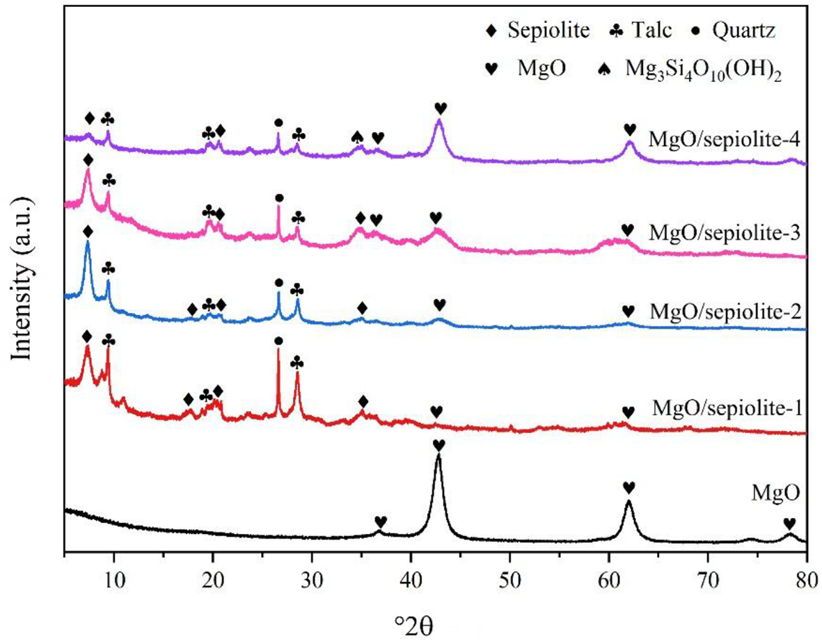
The XRD traces of the MgO/sepiolite composite samples with different nano-MgO loadings are shown in Fig. 1. The diffraction peaks at 36.86°2θ, 42.82°2θ, 62.17°2θ, 74.51°2θ and 78.44°2θ can be assigned to (111), (200), (220), (311) and (222) planes, respectively, of MgO nanocrystals with a periclase phase structure (Singh et al., Reference Singh, Singh, Shukla, Singh and Tandon2016). As shown in Fig. S1, the intensity of the characteristic diffraction peak corresponding to the (110) crystal plane of sepiolite, as well as the quartz diffraction peak, gradually decreased with increasing HCl concentration. When the HCl concentration reached 2.4 mol L–1, the characteristic diffraction peaks of sepiolite nearly disappeared, along with the appearance of a broad diffuse peak characteristic of amorphous SiO2, which indicates that the high-concentration HCl partially destroyed the Mg–O octahedral of the sepiolite framework structure (Wang et al., Reference Wang, Dong, Tian, Sun, Wang and Hui2019), leading to an increased concertation of leached Mg2+ ions (Fig. S2). Through comparison with Fig. S1, it can be seen that the MgO/sepiolite composite exhibited characteristic MgO diffraction peaks at 42.82°2θ and 62.16°2θ (Fig. 1), confirming that the Mg2+ ions released from sepiolite during acid treatment were subsequently captured on the sepiolite nanofibre and converted into MgO NPs via the hydrothermal reaction. In Fig. 1, the most significant-intensity diffraction peak at 42.82°2θ was assigned to the (200) crystal plane of MgO, suggesting preferential growth along this direction (Sidhu et al., Reference Sidhu, Bala, Singh, Ahuja and Kumar2020). Furthermore, the intensity of the MgO characteristic peaks increased with the amount of leached Mg2+, indicating that the loading of MgO NPs on sepiolite increased accordingly (Table S2).
XRD traces of MgO and the MgO/sepiolite (Sep) composites.

Figure 1 Long description
Intensity (a.u) Five stacked line traces are shown, each labeled at the right end: MgO, MgO/sepiolite-1, MgO/sepiolite-2, MgO/sepiolite-3 and MgO/sepiolite-4. A legend at the top lists symbol markers for phases: Sepiolite, MgO, Talc, Mg2Si4O11(OH)2 and Quartz. The x-axis is labeled 2theta, ranging from 10 to 80 with tick marks at 10, 20, 30, 40, 50, 60, 70 and 80. The y-axis is labeled Intensity (a.u) with no numeric tick labels. MgO trace: a low baseline with three prominent narrow peaks, including a tall peak near 43 on the 2theta axis and additional peaks near about 62 and about 75. MgO/sepiolite-1 trace: multiple narrow peaks across the range, including peaks near about 19, about 30, about 37, about 43, about 62 and about 75. MgO/sepiolite-2 trace: multiple narrow peaks across the range, including peaks near about 19, about 30, about 37, about 43, about 62 and about 75. MgO/sepiolite-3 trace: multiple narrow peaks across the range, including peaks near about 19, about 30, about 37, about 43, about 62 and about 75. MgO/sepiolite-4 trace: multiple narrow peaks across the range, including peaks near about 19, about 30, about 37, about 43, about 62 and about 75. Symbol markers are placed above many peaks on the composite traces, matching the legend categories.
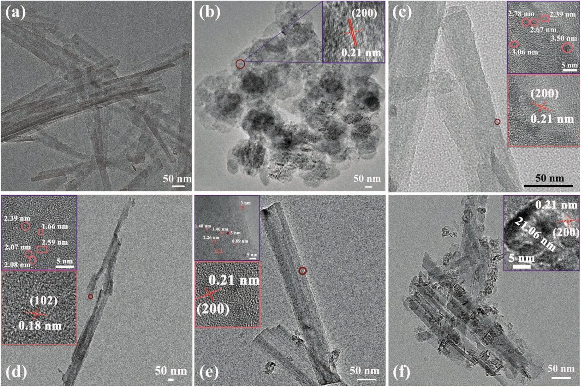
Figure 2 shows TEM images of MgO/sepiolite composites with different nano-MgO loadings obtained through the acid leaching–hydrothermal combined reaction. After treatment with 1.2 mol L–1 HCl (Fig. 2a, b), the sepiolite retained its characteristic nanofibrous morphology, while the MgO NPs exhibited a sheet-like morphology arranged in stacked sheets, with individual particle sizes of ∼100–130 nm and clear agglomeration. Notably, when Mg2+ ions were leached from sepiolite and subsequently loaded onto the fibres to form MgO NPs (Fig. 2c–f), the particle size decreased significantly. This result confirms that sepiolite effectively inhibited the nucleation and growth of MgO NPs. Specifically, after treatment with 0.12 mol L–1 HCl followed by hydrothermal treatment, the sepiolite structure showed negligible change, with little Mg2+ leaching detected (Fig. S2). Consequently, the sepiolite surface exhibited only a low-density distribution of MgO NPs, with particle sizes of ∼2–3 nm (Fig. 2c). When the HCl concentration increased to 1.2 mol L–1 (Fig. 2d), MgO NPs with a uniform size of 1.0–2.5 nm were loaded onto the sepiolite fibre surface with high density and excellent dispersion (Fig. S3). Upon further increasing the HCl concentration to 2.4 mol L–1 (Fig. 2e), the sepiolite fibre surface became roughened due to excessive leaching of Mg2+ ions. In addition, the high density of MgO NPs formed on the sepiolite surface exhibited pronounced agglomeration, with particle sizes correspondingly increasing to ∼20 nm.
TEM images of (a) sepiolite treated with acid, (b) MgO, (c) MgO/sepiolite-1, (d) MgO/sepiolite-2, (e) MgO/sepiolite-3 and (f) MgO/sepiolite-4.

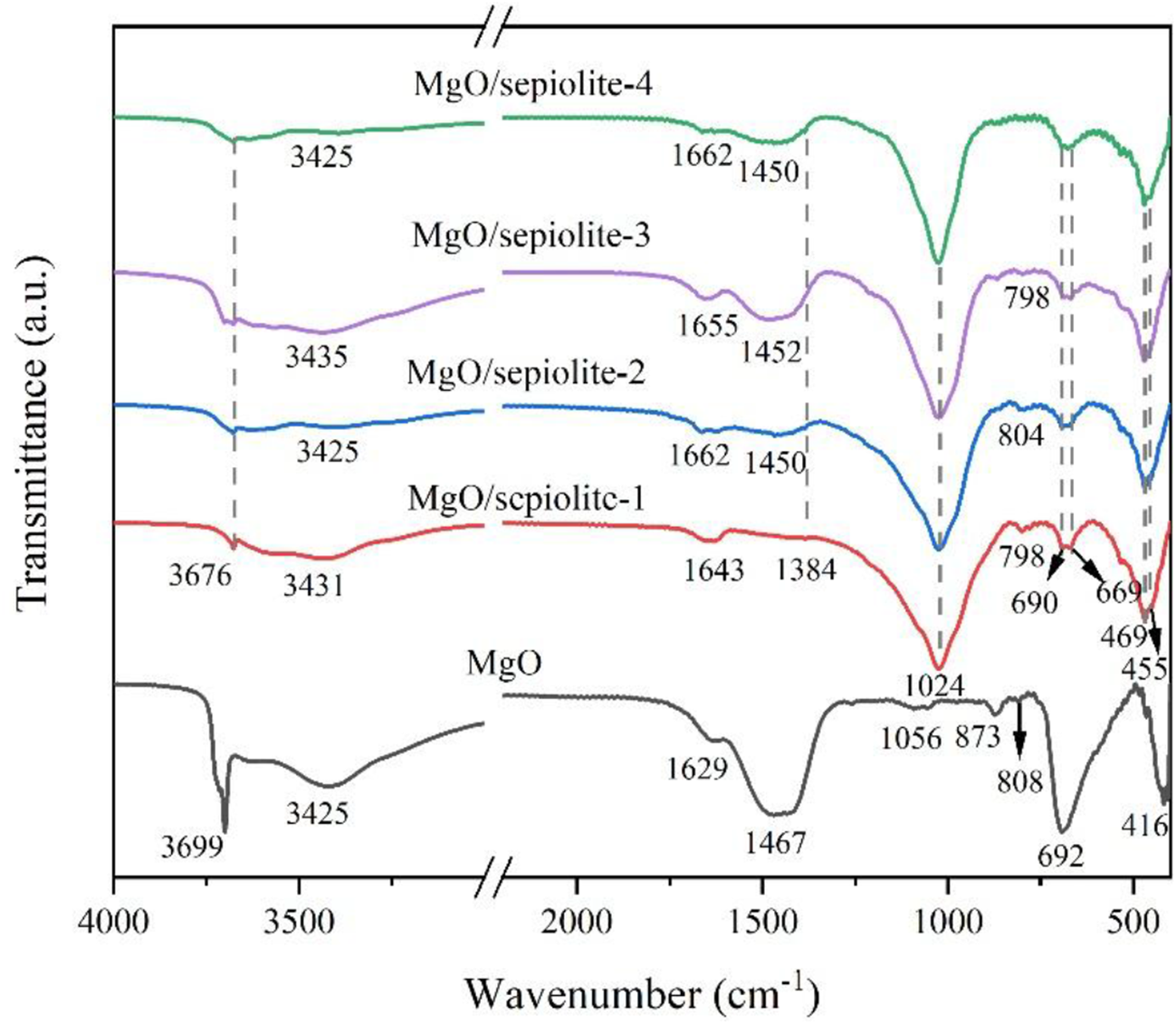
To further investigate the surface functional groups and bonding states of the MgO/sepiolite composites, FTIR spectra were carried out on sepiolite, acid-treated sepiolite and the MgO/sepiolite composites (Figs 3 & S4). As shown in Fig. S4, after the acid leaching reaction, the band at 3675 cm–1 assigned to the hydroxyl groups (Mg3OH) coordinated with octahedra Mg2+ in sepiolite disappeared, indicating that Mg2+ ions were leached from sepiolite. In addition, a slight increase in the intensity of the Si–O–Si band at 468 cm–1 was observed, which indicates the generation of more Si–O dangling bonds and Si–O broken bonds due to the leaching of Mg2+ ions from the Si–O–Mg–O–Si bonds (Li et al., Reference Li, Tian, Chen and Liang2022b). Additionally, the intensity of the bands at 3564 and 1668 cm–1 increased, suggesting that the sepiolite structure was not completely destroyed after acid treatment. In Fig. 3, the bands at 3425 and 1629 cm–1 correspond to the stretching and bending vibrations of hydroxyl groups adsorbed on the MgO surface, respectively (Pandiselvi et al., Reference Pandiselvi, Manikumar and Thambidurai2014; Karthik et al., Reference Karthik, Dhanuskodi, Gobinath, Prabukumar and Sivaramakrishnan2019). The bands at 3699 and 416 cm–1 are related to the symmetric stretching vibration of the Mg–OH bond and the stretching vibration of the Mg–O bond, respectively (Karthik et al., Reference Karthik, Dhanuskodi, Gobinath, Prabukumar and Sivaramakrishnan2019). Additionally, the new band at 1384 cm–1 is assigned to the carbonate species (CO32–) adsorbed on the MgO/sepiolite composite surface during preparation, whereas the band at 455 cm–1 is attributed to the stretching vibration of the Mg–O bond in the MgO/sepiolite composites. Compared with sepiolite (Fig. S4), the band of the Si–O–Si bonds of the acid-treated sepiolite shifted from 1026 cm–1 to a lower wavenumber, indicating that the formation of weak Si–O–Mg bonds within the composite structure (Ou et al., Reference Ou, Zhou, Zhao, Geng, Hao and Xu2012). These results demonstrated that the Mg2+ ions leached from sepiolite were successfully transformed into MgO NPs and loaded onto the sepiolite fibre via the acid leaching–hydrothermal combined reaction.
FTIR spectra of MgO and the MgO/sepiolite (Sep) composites.

Figure 3 Long description
Stacked line plot titled “MgO/sepiolite-4”, “MgO/sepiolite-3”, “MgO/sepiolite-2”, “MgO/sepiolite-1” and “MgO”. Y-axis label: Transmittance (a.u.). X-axis label: Wavenumber (cm superscript minus 1). Major ticks shown at 4000, 3500, 2000, 1500, 1000 and 500, with a broken-axis mark between the 3500 region and the 2000 region. Five spectra are shown, each labeled next to its line. MgO/sepiolite-4 spectrum: labeled bands at 3425, 1662, 1450, 798, 690, 469 and 455. MgO/sepiolite-3 spectrum: labeled bands at 3435, 1655, 1452 and 804. MgO/sepiolite-2 spectrum: labeled bands at 3425, 1662, 1450, 798, 690, 469 and 455. MgO/sepiolite-1 spectrum: labeled bands at 3676, 3431, 1643, 1384, 1056, 873, 808, 798, 692, 469, 455 and 416. MgO spectrum: labeled bands at 3699, 3425, 1629 and 1467.
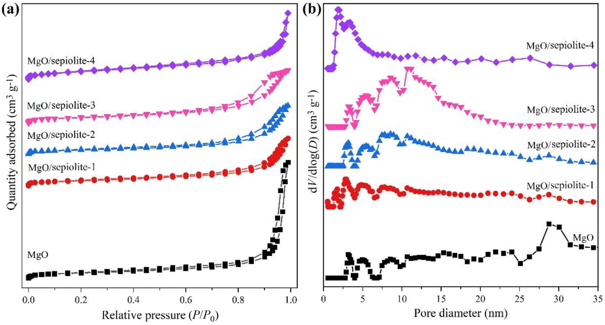
The specific surface area and pore properties of the MgO/sepiolite composites were further investigated by N2 adsorption–desorption experiments, and the results are shown in Fig. 4a, b. The MgO displayed a Type-II isotherm and an H3 hysteresis loop, suggesting that the stacked MgO nanosheets formed abundant crevice and crack pores (Thommes & Cychosz, Reference Thommes and Cychosz2017). In addition, all MgO/sepiolite composites exhibited Type-IV isotherms and H3 hysteresis loops, confirming the presence of mesoporous structures (Fig. 4a). As shown in Fig. 4b, the corresponding pore-size distributions of the MgO/sepiolite composites were mainly in the range of 3–15 nm, further demonstrating their mesoporous characteristics. The pore structure was further analysed by comparing the pore-size distributions of sepiolite and acid-treated sepiolite (Fig. S5) and the MgO/sepiolite composites. Acid treatment of sepiolite resulted in an increased micropore content because carbonate impurities on the sepiolite fibres reacted with HCl, which exposed previously blocked pores, including pores parallel and perpendicular to the fibre axis (Tang et al., Reference Tang, Wang, Tang, Liang and Ren2012). With increasing HCl concentration, the leaching of Mg2+ from the sepiolite led to the connection of internal channels within the crystal, and some micropores expanded into mesopores. When most of the Mg2+ was removed, some micropores and mesopores further expanded into macropores. The amount of loaded MgO increased with HCl concentration, as a higher acid concentration leached more Mg2+, which was subsequently converted to MgO NPs via the hydrothermal reaction. When the HCl concentration reached 2.4 mol L–1, most of Mg2+ was leached, resulting in the highest MgO loading (MgO/sepiolite-4). Consequently, with increasing MgO loading, the micropores of the MgO/sepiolite composites decreased while the mesopores increased. Moreover, higher MgO loading caused agglomeration and larger MgO particles, which generated interparticle pores on the sepiolite surface (Fan et al., Reference Fan, Yuan, Bergaya, Chen, Zhu and Liu2011).
(a) N2 adsorption–desorption isotherm and (b) pore-size distribution of MgO and the MgO/sepiolite composites.

Figure 4 Long description
Line plot with multiple series. The x-axis label is Relative pressure (P slash P0), ranging from 0.0 to 1.0 with labeled ticks at 0.0, 0.2, 0.4, 0.6, 0.8 and 1.0. The y-axis label is Quantity adsorbed (cm superscript 3 g superscript -1). Five labeled series appear: MgO, MgO slash sepiolite-1, MgO slash sepiolite-2, MgO slash sepiolite-3 and MgO slash sepiolite-4. Each series increases gradually across most of the x-range and then rises steeply near relative pressure close to 1.0. Some series show paired traces near the high-pressure end. Line plot with multiple series. The x-axis label is Pore diameter (nm), ranging from 0 to 35 with labeled ticks at 0, 5, 10, 15, 20, 25, 30 and 35. The y-axis label is dV slash dD (cm superscript 3 g superscript -1). Five labeled series appear: MgO, MgO slash sepiolite-1, MgO slash sepiolite-2, MgO slash sepiolite-3 and MgO slash sepiolite-4. The curves show higher values at smaller pore diameters and then generally decrease and level out as pore diameter increases, with local peaks and fluctuations across the range.
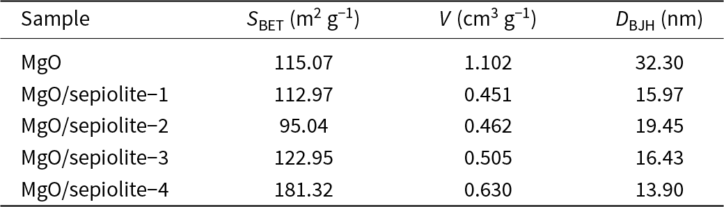
The surface parameters of MgO and the MgO/sepiolite composites are summarized in Table 1. As Mg2+ ions were leached from sepiolite and subsequently loaded onto its surface as MgO NPs, the specific surface area of the composites initially decreased from 112.97 to 95.04 m2 g–1 with increasing MgO loading. This result was attributed to the fact that the MgO NPs loaded onto the sepiolite surface partially blocked its pores during the synthesis process, which significantly reduced the specific surface area of the MgO/sepiolite composites. However, upon further increasing the loading amount (MgO/sepiolite-3 and MgO/sepiolite-4), the specific surface area increased again from 122.95 to 181.32 m2 g–1. This was caused by the nano-sized MgO particles loaded on the sepiolite surface, which provided an additional specific surface area due to their small particle size and excellent dispersion, thereby mitigating the reduction and MgO/sepiolite composites and ultimately increasing the overall specific surface area. In addition, as the HCl concentration increased, the effective dissolution of impurities such as calcium carbonate reduced the binding between sepiolite fibre bundles, consequently increasing the specific surface area (Wu et al., Reference Wu, Hong, Zhan, Tang, Cui and Kou2022).
Pore structural characteristics of MgO and MgO/sepiolite composites.

Table 1 Long description
Pore structure metrics are listed for MgO and four MgO sepiolite composites, including specific surface area, total pore volume, and average pore size. Pure MgO has a surface area of 115.07 square meters per gram, pore volume 1.102 cubic centimeters per gram, and average pore size 32.30 nanometers. Among composites, MgO/sepiolite-4 stands out with the highest surface area at 181.32 square meters per gram and the smallest average pore size at 13.90 nanometers, with pore volume 0.630 cubic centimeters per gram. MgO/sepiolite-1 through -3 have surface areas from 95.04 to 122.95 square meters per gram and pore volumes clustered near 0.451 to 0.505 cubic centimeters per gram, with average pore sizes from 15.97 to 19.45 nanometers. Compared with MgO, all composites show much lower pore volume but smaller average pore sizes. Values are descriptive and do not indicate measurement uncertainty or statistical significance.
D BJH = average pore size; S BET = specific surface area; V = total pore volume.
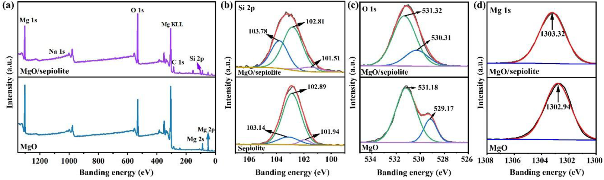
The surface element composition and valence states of the sepiolite, MgO and MgO/sepiolite composites were further investigated by XPS analysis, and the results are shown in Fig. 5. As presented in Fig. 5a, the survey spectrum of the MgO/sepiolite composite showed that Si, O and Mg were the main compositional elements. Furthermore, the intensity of Mg elements changed compared to that sepiolite, suggesting that the chemical state of Mg element was altered during the synthesis of the MgO/sepiolite composite. High-resolution XPS spectra of Si 2p for sepiolite and the MgO/sepiolite composite are shown in Fig. 5b. The peaks at 101.51, 102.81 and 103.78 eV were related to Si–OH, Si–O–Mg and Si–O–Si, respectively. Following the leaching of Mg2+ ions and subsequent formation of MgO NPs on the sepiolite fibre, the peak area ratio of Si–O–Si increased, whereas that of Si–O–Mg decreased. This change can be attributed to the acid treatment of sepiolite, which led to the leaching of Mg2+ ions from the sepiolite structure and the generation of additional Si–O bonds. In addition, the incomplete re-immobilization of leached Mg2+ ions onto the sepiolite surface for MgO formation further contributed to the changes in the Si–O–Mg and Si–O–Si area ratios. The O 1s high-resolution XPS spectrum of MgO (Fig. 5c) exhibits two peaks at 529.17 and 531.18 eV, corresponding to lattice oxygen and adsorbed oxygen (oxygen chemically adsorbed in oxygen vacancies), respectively (Veldurthi et al., Reference Veldurthi, Shin, Joo and Jung2012). Compared with MgO, the MgO/sepiolite composite exhibited a significant increase in adsorbed oxygen content, confirming the formation of additional oxygen vacancies that promote reactive oxygen species (ROS) generation and antimicrobial activity. Additionally, the O 1s peak shifted towards higher energy, which is attributed to the coordination of Mg2+ ions with the sepiolite (Cheng et al., Reference Cheng, Wang, Li, Gao, Gao and Guo2024). As shown in Fig. 5d, the Mg 1s peak shifted towards lower binding energies, reflecting a change in the coordination environment of Mg due to interfacial interactions between MgO and sepiolite (Hui et al., Reference Hui, Yan, Mu, Kang, Zhou and Wang2020).
XPS spectra of sepiolite, MgO and the MgO/sepiolite-3 composite: (a) survey, (b) Si 2p, (c) O 1s and (d) Mg 1s.

Figure 5 Long description
A) Survey X ray photoelectron spectroscopy spectrum comparing three offset line spectra labeled MgO/sepiolite, sepiolite and MgO. X-axis: Binding energy (eV), 0 to 1200. Y-axis: Intensity (a.u.). The MgO/sepiolite spectrum has labeled peaks Mg 1s, O 1s, Mg KLL, C 1s, Si 2p and Na 1s. The plot presents element-labeled peak positions as a comparison across the three spectra. B) High-resolution Si 2p X ray photoelectron spectroscopy line plot with peak fitting for two offset spectra labeled MgO/sepiolite and sepiolite. X-axis: Binding energy (eV), 96 to 106. Y-axis: Intensity (a.u.). Each spectrum shows a black measured curve, a red fitted envelope and multiple fitted component curves. Labeled component peak positions on MgO/sepiolite are 101.51, 102.81 and 103.78. Labeled component peak positions on sepiolite are 101.94, 102.89 and 103.61. The labeled peak positions differ between the two spectra. C) High-resolution O 1s X ray photoelectron spectroscopy line plot with peak fitting for two offset spectra labeled MgO/sepiolite and MgO. X-axis: Binding energy (eV), 524 to 536. Y-axis: Intensity (a.u.). Each spectrum shows a black measured curve, a red fitted envelope and two fitted component curves. Labeled component peak positions on MgO/sepiolite are 531.32 and 529.30. Labeled component peak positions on MgO are 531.18 and 529.17. The labeled peak positions differ between the two spectra. D) High-resolution Mg 1s X ray photoelectron spectroscopy line plot for two offset spectra labeled MgO/sepiolite and MgO. X-axis: Binding energy (eV), 1302 to 1308. Y-axis: Intensity (a.u.). Each spectrum shows a black measured curve and a red fitted envelope. The labeled peak position on MgO/sepiolite is 1303.33. The labeled peak position on MgO is 1302.43. The labeled peak position differs between the two spectra.
Based on these results, a possible synthesis mechanism for the MgO/sepiolite composites is proposed. Firstly, as the HCl concertation increased, the Mg–O bonds within the octahedral structure of sepiolite were gradually attacked and broken by H+ ions, leading to the release of free Mg2+ ions (Abdullayev et al., Reference Abdullayev, Joshi, Wei, Zhao and Lvov2012). Owing to the high bond energy, the Si–O bonds were difficult to break under the attack of H+ ions. Consequently, the leaching of Mg2+ ions from Si–O–Mg–O–Si bonds generated coordination vacancies (Li et al., Reference Li, Tian, Chen and Liang2022b), with the formation of Si–O dangling bonds or Si–O broken bonds at the residual sites (Equation 1). To balance the charge, the reactive Si–O dangling bonds and Si–O broken bonds on the Si–O tetrahedra of sepiolite combined with H+ ions in the solution to form Si–O–H groups (Valentín et al., Reference Valentín, López-Manchado, Posadas, Rodríguez, Marcos-Fernández and Ibarra2006; Li et al., Reference Li, Nie, Tang, Tian and Liang2022a). However, Si–OH bonds are susceptible to hydrolysis in aqueous solution, thus forming Si–O– and H⁺ (Equation 2). Subsequently, the addition of NaOH provided a large amount of OH– ions, which preferentially reacted with the more reactive Mg2+ ions in the solution, resulting in a large quantity of Mg(OH)+ (Equations 3 & 4). Both Mg2+ and Mg(OH)+ ions exhibited a strong affinity for the negatively charged Si–O– groups on the sepiolite surface, leading to neutralization reactions that resulted in the deposition of Mg(OH)2 (Equation 5). During the subsequent hydrothermal reaction, the nuclei gradually aggregated and grew into Mg(OH)2 NPs. Finally, the Mg(OH)2 was transformed into MgO NPs via dehydration upon calcination (Equation 6), successfully forming the MgO/sepiolite composites.
 \begin{equation}{\text{Si--O\ dangling bonds or Si--O broken bonds + }}{{\text{H}}^{\text{+ }}} \to {\text{Si--OH}}\end{equation}
 \begin{equation}{\text{Si--OH}} \to {\text{Si--}}{{\text{O}}^{\text{--}}}{\text{ + }}{{\text{H}}^{\text{+ }}}\end{equation}
 \begin{equation}{\text{NaOH}} \to {\text{N}}{{\text{a}}^{\text{+ }}}{\text{ + O}}{{\text{H}}^{{\text{-- }}}}\end{equation}
 \begin{equation}{\text{M}}{{\text{g}}^{{\text{2+ }}}}{\text{ + O}}{{\text{H}}^{\text{--}}} \to {\text{Mg}}{\left( {{\text{OH}}} \right)^{\text{+ }}}\end{equation}
 \begin{equation}{\text{Mg}}{\left( {{\text{OH}}} \right)^{\text{+ }}}{\text{ + O}}{{\text{H}}^{\text{--}}} \to {\text{Mg}}{\left( {{\text{OH}}} \right)_{\text{2}}}\end{equation}
 \begin{equation}{\text{Mg}}{\left( {{\text{OH}}} \right)_{\text{2}}}{ }\mathop \to \limits^\Delta {\text{MgO + }}{{\text{H}}_{\text{2}}}{\text{O}}\end{equation}
Antimicrobial activity
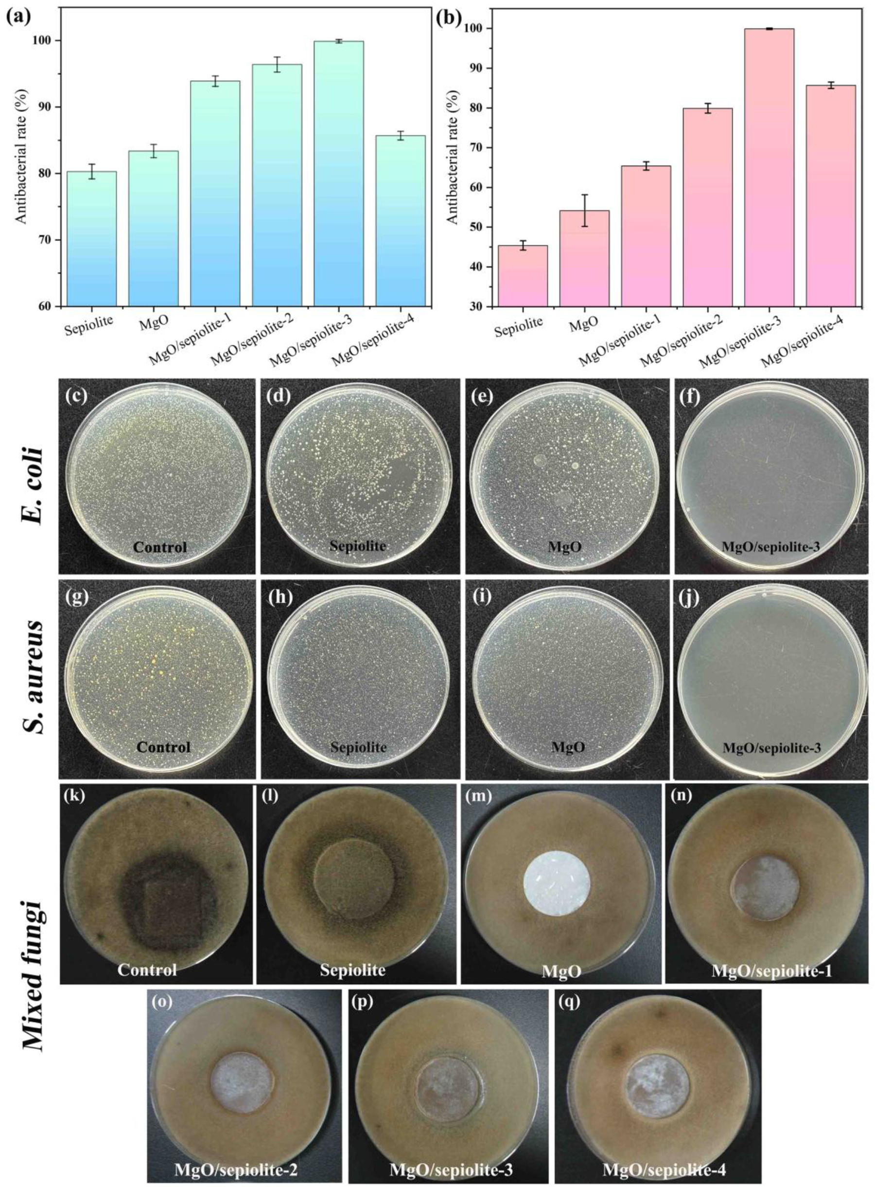
The antibacterial activity of sepiolite, MgO and the MgO/sepiolite composites was investigated against E. coli and S. aureus, and the results are shown in Fig. 6. The antibacterial rates of sepiolite were 80.30% against E. coli and 83.38% against S. aureus, while MgO showed antibacterial rates of 45.40% and 54.18%, respectively (Fig. 6a, b). The antibacterial activity of MgO/sepiolite composites progressively improved with increasing HCl concertation. These results were further supported by bacteria plate cultures, as shown in Fig. 6c–j. Among the composites, MgO/sepiolite-3 showed the best antibacterial performance, with a 99.99% antibacterial rate against both E. coli and S. aureus, whereas MgO/sepiolite-4 showed antibacterial rates below 90.00% against both bacterial strains. The antibacterial performance was closely associated with the loading amount and dispersion of MgO NPs on the sepiolite fibre surface. For MgO/sepiolite-4, the excessive loading of MgO NPs caused agglomeration on the sepiolite surface, which reduced both the specific surface area and the antibacterial active sites, thereby limiting its antibacterial performance. In addition, the MgO/sepiolite composites exhibited better antibacterial performance against E. coli than against S. aureus. This difference can be attributed to the structural characteristics of the bacterial cell walls. Gram-negative bacteria such as E. coli have thinner cell walls that lack the three-dimensional structure formed by the tetrapeptide side chains cross-linked via pentapeptide cross-bridges, which is a structure typical of the peptidoglycan layer in bacteria such as S. aureus (Leus et al., Reference Leus, Adamiak, Chandar, Bonifay, Zhao and Walker2023; Zhang et al., Reference Zhang, Su, Chen, Qiao, Yang and Zhao2023). Consequently, Gram-negative bacterial cell walls provide less effective protection for their internal metabolic processes, making them more prone to attack by antimicrobial materials.
Antimicrobial properties of sepiolite, MgO and the MgO/sepiolite composites. The antimicrobial rate for (a) E. coli and (b) S. aureus and the antimicrobial activity for (c–f) E. coli, (g–j) S. aureus and (k–q) mixed fungi.

Figure 6 Long description
This multi-panel scientific figure includes bar charts and petri dish images. The top row features bar charts showing antimicrobial rates. Chart (a) displays rates against E. coli, with sepiolite at approximately 80 percent, MgO at 45 percent and MgO/sepiolite-3 reaching nearly 100 percent. Chart (b) shows rates against S. aureus, with sepiolite at about 83 percent, MgO at 54 percent and MgO/sepiolite-3 also near 100 percent. Below, petri dish images illustrate bacterial growth. For E. coli (c-f), control shows dense colonies, while MgO/sepiolite-3 shows clear inhibition. For S. aureus (g-j), similar trends are observed. Mixed fungi images (k-q) show varying inhibition, with MgO/sepiolite-3 performing best. The layout follows a top-to-bottom reading order, starting with bar charts, then E. coli, S. aureus and mixed fungi sections. The key takeaway is that MgO/sepiolite-3 exhibits the highest antimicrobial performance across all tested organisms.
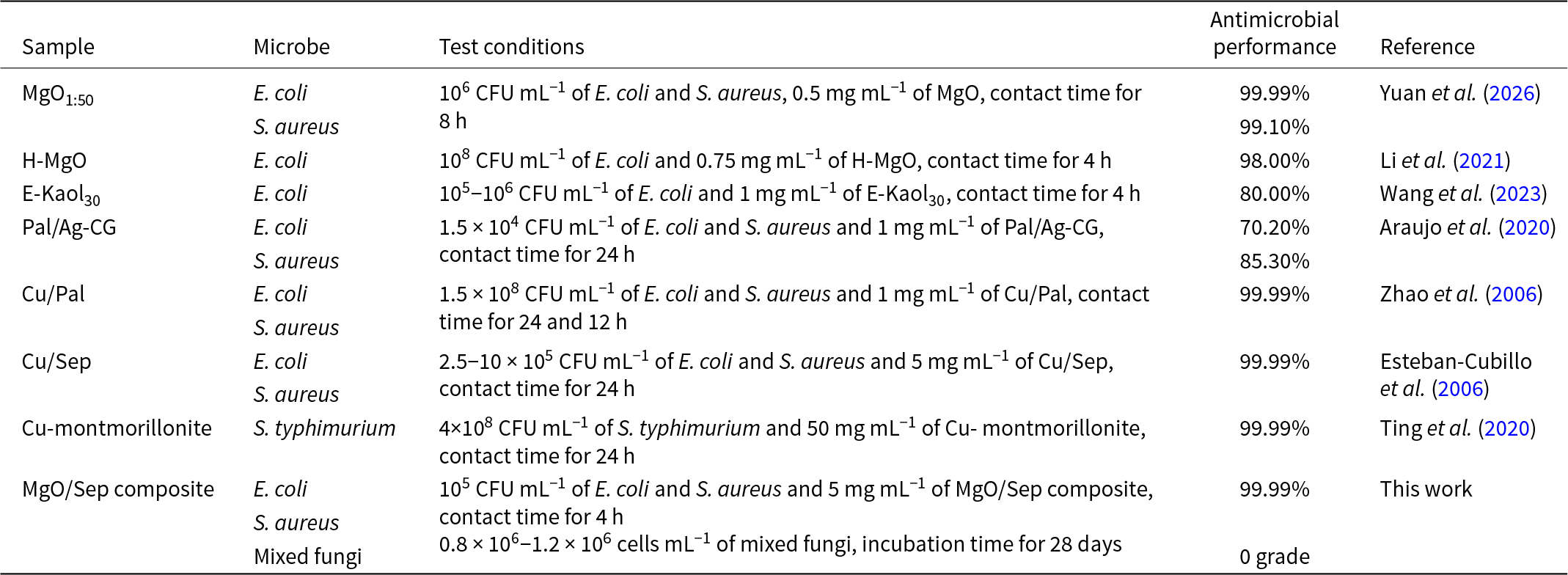
The antifungal activity of sepiolite, MgO and the MgO/sepiolite composites against mixed fungi was also evaluated, with the results shown in Fig. 6k–q. After 28 days of incubation, the control group and sepiolite sample were completely covered with fungi, corresponding to a grade 4 fungal growth level. All MgO/sepiolite composites exhibited a grade 0 fungal growth level, indicating excellent antifungal performance. Further observation of fungal growth in the samples revealed that MgO/sepiolite-3 exhibited the most effective antifungal activity. This superior performance is ascribed to its beneficial MgO loading amount and the uniform distribution of MgO NPs on the sepiolite fibre surface. Consistent with the antibacterial results, MgO/sepiolite-3 exhibited the best antimicrobial performance against E. coli, S. aureus and mixed fungi. These analyses indicate that the MgO/sepiolite composites possess excellent broad-spectrum antibacterial properties. Table 2 summarizes the antimicrobial performance of previously reported MgO and other clay-based antimicrobial agents. In comparison, MgO/sepiolite-3 exhibited excellent antimicrobial performance.
Comparative antimicrobial performance of as-prepared samples and other clay-based antimicrobial agents reported previously.

Table 2 Long description
Comparative results list antimicrobial performance for multiple clay-based or mineral-based samples against specific microbes under stated inoculum levels, dosages, and contact times. Several materials report 99.99% reduction for bacteria: MgO1:50 against E. coli and S. aureus after eight hours; Cu/Pal against E. coli (and also tested on S. aureus) with contact times of twelve to twenty-four hours; Cu/Sep against E. coli and S. aureus after twenty-four hours; Cu-montmorillonite against S. typhimurium after twenty-four hours; and the MgO/Sep composite from this work against E. coli and S. aureus after four hours. Lower bacterial reductions are reported for H-MgO against E. coli at 98.00% after four hours and for E-Kaol30 against E. coli at 80.00% after four hours. Pal/Ag-CG shows moderate performance, with 70.20% for E. coli and 85.30% for S. aureus after twenty-four hours. Only one entry reports a non-bacterial outcome: the MgO/Sep composite is rated “0 grade” against mixed fungi after a long incubation period. Comparisons should be interpreted cautiously because starting microbe concentrations, material doses, and exposure times vary across studies.
Ag-CG = silver nanoparticles stabilized with cashew gum; CFU = colony-forming unit; Kaol = kaolinite; Pal = palygorskite; Sep = sepiolite.
Antimicrobial mechanism of the nano-MgO/sepiolite composites
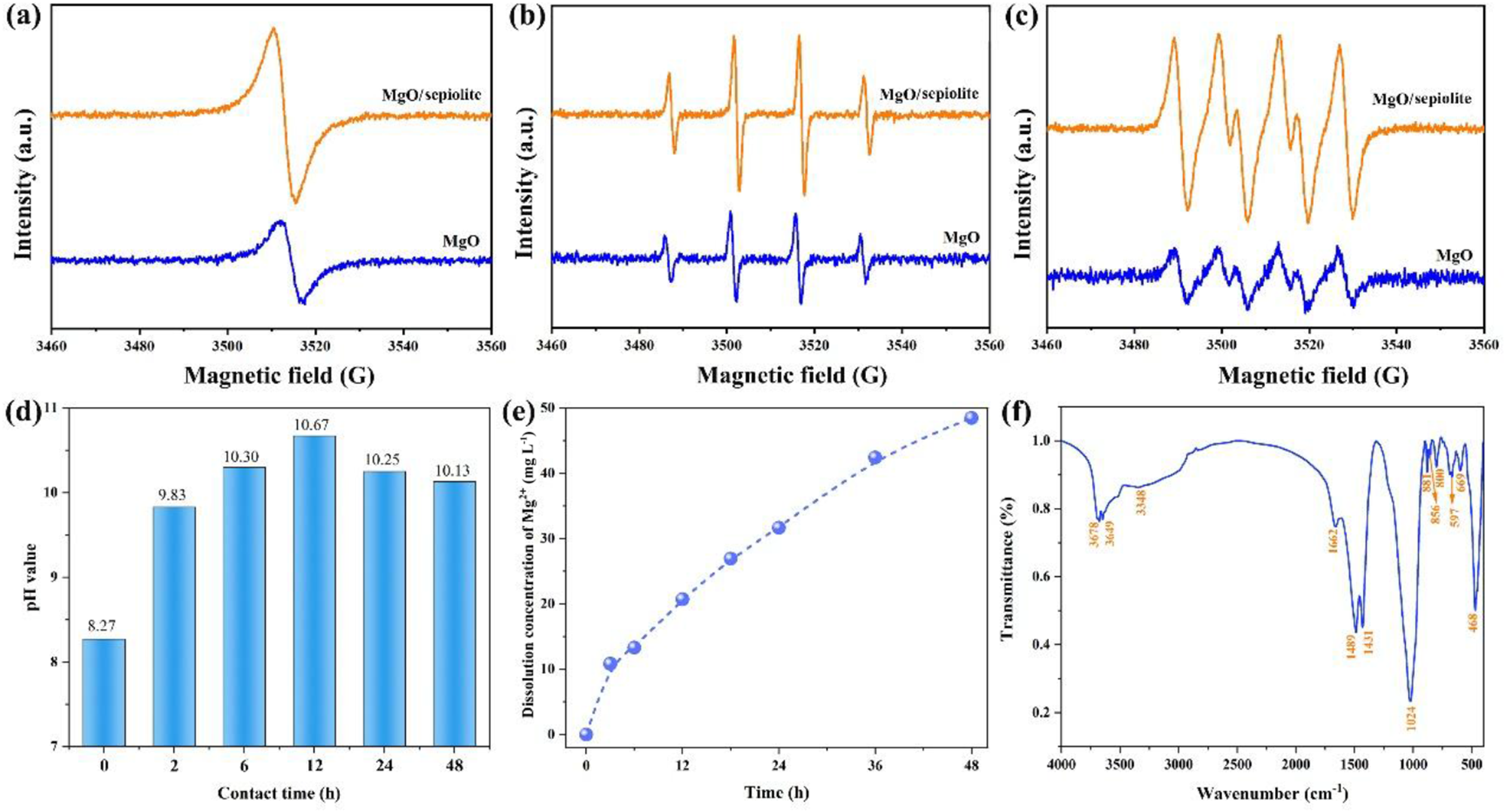
ESR analysis was employed to explore the role of oxygen vacancies and ROS (·OH and ·O2–) generation in the antimicrobial mechanism of MgO and the MgO/sepiolite composites, and the results are shown in Fig. 7a–c. When ROS are present in the environment, they can disrupt the microbial cell wall, leading to the leakage of intracellular contents, which directly results in microbial death (Rajeshkumar & Bharath, Reference Rajeshkumar and Bharath2017; Khezerlou et al., Reference Khezerlou, Alizadeh-Sani, Azizi-Lalabadi and Ehsani2020). Once the cell wall is disrupted, these radicals can more readily penetrate the cell interior, where they interact with essential cellular component such as proteins, ribosomes and genetic material (Nguyen et al., Reference Nguyen, Nguyen, Nguyen, Nguyen, Nguyen and Tran2022), thereby causing oxidative damage, leading to microbial death. As shown in Fig. 7a, the MgO/sepiolite-3 composite exhibited a significantly higher oxygen vacancy intensity than MgO, indicating that its surface possessed numerous defects and abundant oxygen vacancies. These features indicate a potential pathway for oxygen vacancy-mediated ROS damage to microorganisms. Furthermore, the characteristic signals corresponding to ·OH and ·O2– radicals were detected in both MgO and MgO/sepiolite-3 (Fig. 7b, c). These results suggests that oxygen vacancies act as electron donors, which react with water and oxygen in the environment through electron reduction reactions, thereby generating ·O2– (Equations 7 & 8); subsequently, ·O2– can be converted into H2O2 and ·OH through a series of reactions (Equations 9–11; Li et al., Reference Li, Hong, Yang, Zhao, Diko and Zhu2021; Biswas et al., Reference Biswas, Kar and Jana2022). Moreover, MgO/sepiolite-3 generated greater amounts of ·O2– and ·OH radicals compared to MgO, which is related to the number of oxygen vacancies in the sample, as discussed previously. Importantly, the greater oxygen vacancy concentration in MgO/sepiolite-3 is closely correlated with the particle size of MgO and the specific structural of sepiolite. A smaller MgO particle size results in more oxygen vacancies, while acid treatment also introduced oxygen vacancies on the sepiolite surface. Moreover, the abundant Si–O–Si bonds in sepiolite can break during composite preparation, possibly generating reactive species such as ≡Si· or ≡SiO· radicals. These silicon-based radical species can subsequently react with H2O or O2, contributing to the generation of additional ·OH radicals (Equations 12–14; Narayanasamy & Kubicki, Reference Narayanasamy and Kubicki2005; He et al., Reference He, Wu, Zhu, Lin, Lv and Xian2023).
 \begin{equation}{{\text{H}}_{\text{2}}}{\text{O}} \to {\text{O}}{{\text{H}}^{\text{--}}}{\text{ + }}{{\text{H}}^{\text{+ }}}\end{equation}
 \begin{equation}{{\text{O}}_{\text{v}}}\left( {{{\text{e}}^{\text{--}}}} \right){\text{ + }}{{\text{O}}_{\text{2}}} \to \cdot {{\text{O}}_{\text{2}}}^{\text{--}}\end{equation}
 \begin{equation} \cdot {{\text{O}}_{\text{2}}}^{\text{--}}{\text{ + O}}{{\text{H}}^{\text{--}}} \to \cdot {\text{OH + }}{{\text{O}}_{\text{2}}}^{{\text{2--}}}\end{equation}
 \begin{equation}{{\text{O}}_{\text{2}}}^{{\text{2--}}}{\text{ + 2}}{{\text{H}}_{\text{2}}}{\text{O}} \to {{\text{H}}_{\text{2}}}{{\text{O}}_{\text{2}}}{\text{ + 2O}}{{\text{H}}^{\text{--}}}\end{equation}
 \begin{equation}{{\text{H}}_{\text{2}}}{{\text{O}}_{\text{2}}}{\text{ + }}{{\text{O}}_{\text{v}}}\left( {{{\text{e}}^{\text{--}}}} \right) \to\cdot {\text{OH + O}}{{\text{H}}^{\text{--}}}\end{equation}
 \begin{equation} \equiv\!{\text{Si}\!\cdot + }{{\text{O}}_{\text{2}}} \to \equiv\!{\text{SiOO}}\cdot\end{equation}
 \begin{equation} \equiv\!{\text{SiOO}}\!\cdot {\text{ + }}{{\text{H}}_{\text{2}}}{\text{O }} \to \equiv\!{\text{SiOOH + }} \cdot\!{\text{OH}}\end{equation}
 \begin{equation} \equiv\!{\text{SiO}}\!\cdot {\text{ + }}{{\text{H}}_{\text{2}}}{\text{O }} \to \equiv\!{\text{SiOH + }} \cdot\!{\text{OH}}\end{equation}
To further investigate the antimicrobial mechanism of the MgO/sepiolite composites against bacteria and fungi, the pH value changes of the microbial suspensions after contact with the samples was measured. As shown in Fig. 7d, the pH value of the microbial solution increased after contact with the MgO/sepiolite composites, rising from 8.27 to 10.33 within 6 h. This increase is due to the alkaline earth metal oxide characteristic of MgO, which reacts with water molecules to produce OH⁻ ions (MgO + H2O → Mg(OH)⁺ + OH⁻), thereby increasing the pH of the solution (Dong et al., Reference Dong, Cairney, Sun, Maddan, He and Deng2010). Importantly, under alkaline conditions, more OH– are generated, which promotes the reactions shown in Equations 9–11, leading to the increased production of ·OH and thus improving the antimicrobial performance. Additionally, a weakly acidic, humid environment with abundant nutrients at temperatures between 25°C and 30°C is favourable for the rapid growth and proliferation of microorganisms. When the pH value of the growth environment shifts to alkaline conditions, this directly affects the stability and permeability of the cell membrane, the solubility of nutrients and the enzymatic reactions within the microbial cells (Williams, Reference Williams2024).
ESR spectra of MgO and MgO/sepiolite-3 for (a) oxygen vacancy, (b) ·OH and (c) ·O2–. (d) The change in pH values for bacterial solutions (t = 0 h, measured immediately after MgO/sepiolite composite addition). (e) The dissolution concentration of Mg2+ from MgO/sepiolite-3 over time. (f) FTIR spectra of the MgO/sepiolite-3 reaction with the E. coli solution.

Figure 7 Long description
Contains six plots labeled (a) to (f). (a) ESR spectra for oxygen vacancy with intensity (arbitrary units) vs magnetic field (G), showing higher intensity for MgO/sepiolite-3. (b) ESR spectra for hydroxyl radicals, with similar axes, showing distinct peaks for both samples. (c) ESR spectra for superoxide radicals, with similar axes, showing multiple peaks. (d) Bar chart of pH values over contact time (hours), showing an increase from 8.27 to 10.13. (e) Scatter plot of magnesium dissolution concentration over time (hours), showing an upward trend. (f) FTIR spectra with transmittance vs wavenumber (cm superscript -1), showing characteristic peaks for MgO/sepiolite-3.
Figure 7e shows the concentrations of Mg2+ dissolved from the MgO/sepiolite composites at various reaction times. The Mg2+ concentration increased sharply with prolonged reaction time, reaching 31.33 mg L–1 at 24 h and 48.50 mg L–1 at 48 h. These results demonstrated that the MgO/sepiolite composites exhibit excellent Mg2+ release capability, which is closely related to the particle size of the MgO NPs, with smaller particle sizes corresponding to higher Mg2+ dissolution concentrations (Liao et al., Reference Liao, Strayer-Scherer, White, De La Torre-Roche, Ritchie, Colee, Vallad, Freeman, Jones and Paret2019). With the release of Mg2+, the generated magnesium vacancies may cause lattice distortion, which exposes additional oxygen vacancies, increasing the number of active sites for ROS generation. In addition, the dissolution of Mg2+ produces OH⁻, which promotes the conversion of ·O2– to ·OH. Thus, the release of Mg2+ is beneficial to the oxygen vacancy-mediated ROS damaging of microorganisms. Moreover, the strong permeability of Mg2+ and other metal ions facilitates their penetration into microbial cells. Once inside, these ions interfere with the synthesis of essential macromolecules such as proteins and peptides. They can also form chelate complexes with nutrients, impairing microbial metabolism and resulting in cell death (Londono & Williams, Reference Londono and Williams2016). Furthermore, the concentration of Mg2+ dissolved from the MgO/sepiolite composites gradually increased with prolonged reaction time, suggesting that the composites possess excellent sustained-release performance. This is ascribed to the structural characteristics of sepiolite. As a carrier material, it possesses a large specific surface area and abundant pore structure that effectively enhances the sustained release of Mg2+ (Yang & Wang, Reference Yang and Wang2022). Although Mg2+ is released slowly, its sustained release contributes to continuously high antimicrobial activity, making the material suitable for long-term applications.
To gain further insights into the antimicrobial contact-killing mechanism, the FTIR spectra of the MgO/sepiolite-3 composite after contact with E. coli are presented in Fig. 7f. Compared with the MgO/sepiolite-3 composite, two new absorption peaks at 1489 and 1431 cm–1 were observed, which are assigned to the asymmetric and symmetric stretching vibrations of deprotonated –COOH groups from lipopolysaccharides in the E. coli cell wall (Li et al., Reference Li, Feng, Yang, Fu, Wang and Su2010; Leung et al., Reference Leung, Ng, Xu, Shen, Gethings and Wong2014; Pineda et al., Reference Pineda, García-Ruiz, Guaya, Manrique and Osorio2021). The characteristic absorption peak corresponding to the amide groups of lipopolysaccharides appeared at 1662 cm–1 (Kiwi & Nadtochenko, Reference Kiwi and Nadtochenko2005). Moreover, the –OH and Si–O bonds of the MgO/sepiolite-3 composite exhibited changes. These results confirm that E. coli cells were successfully adsorbed onto the surface of the MgO/sepiolite-3 composite, which may be attributed to hydrogen bonding or electrostatic interactions with the anionic carboxyl and phosphate groups of lipopolysaccharides in the E. coli cell wall (Wang et al., Reference Wang, Meng, Jiang, Sarwar, Fu and Yang2023). Therefore, the MgO/sepiolite composites possess an adsorptive affinity for microorganisms, which can increase the contact opportunities between microorganisms and ROS, thus promoting oxygen vacancy-mediated damage to microorganisms. Additionally, the adsorptive interactions potentially modify the cell membrane tension of microorganisms, inducing mechanical deformation and rupture of microbial cells and thus leading to cell death (Rodrigues & Elimelech, Reference Rodrigues and Elimelech2010). Notably, as a natural clay mineral, sepiolite exhibits excellent adsorption performance due to its high specific surface area, nanoporous structure and abundant silanol groups, enabling effective adhesion to microbial cells (Li et al., Reference Li, Tian, Gong, Chen, Kong and Liang2020; Bilgin et al., Reference Bilgin, Bulut and Sabah2023; Bourliva, Reference Bourliva2025).
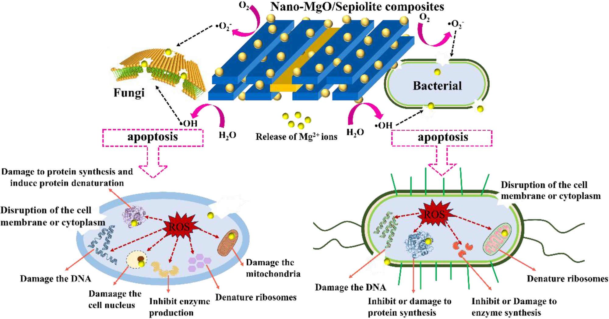
Based on the experimental results and discussion presented herein, a schematic illustration of the proposed antimicrobial mechanism is shown in Fig. 8. These results indicate that that the abundant oxygen vacancies on the MgO/sepiolite composite surface are conducive to generating more ROS and improving the antibacterial ability of these composites. In addition, rapid pH value regulation, Mg2+ release and contact damage were found to play synergistic roles in facilitating oxygen vacancy-mediated antimicrobial damage, resulting in improved antibacterial performance. Consequently, the MgO/sepiolite composites exhibit excellent antibacterial performance.
Illustration of the antimicrobial mechanism of the nano-MgO/sepiolite composites.

Figure 8 Long description
Nano-MgO/sepiolite composites schematic comparing effects on a fungi cell and a bacterial cell, using arrows, dashed boxes and burst icons. A central composite structure is labelled Nano-MgO/Sepiolite composites. Curved arrows around it are labelled O2 minus dot and O2. Text near the arrows includes H2O and OH minus. A label reads Release of Mg2 plus ions. Two dashed boxes contain the word apoptosis, with arrows leading from the central composite area toward each cell. The fungi cell is labelled Fungi. Inside the cell, a burst icon is labelled ROS. Text labels around the fungi cell read: Damage to protein synthesis and induce protein denaturation. Disruption of the cell membrane or cytoplasm. Damage the DNA. Damage the cell nucleus. Damage the mitochondria. Inhibit enzyme production. Denature ribosomes. The bacterial cell is labelled Bacterial. Inside the cell, a burst icon is labelled ROS. Text labels around the bacterial cell read: Disruption of the cell membrane or cytoplasm. Denature ribosomes. Damage the DNA. Inhibit or damage to enzyme synthesis. Inhibit or Damage to protein synthesis.
Conclusion
In summary, nano-MgO/sepiolite composites with excellent antimicrobial performance were successfully synthesized via an acid leaching–hydrothermal combined method using sepiolite as the sole magnesium source. In particular, the MgO/sepiolite-3 composite exhibited synergistic advantages stemming from its fibrous structural characteristics, small MgO particle size (1.0–2.5 nm), large specific surface area (122.95 m2 g–1), high porosity and abundant oxygen vacancies. As a result, this composite achieved high antimicrobial rates of 99.99% against both E. coli and S. aureus and a fungal growth grade of 0. This excellent antimicrobial performance is primarily attributed to the fibrous morphology of the composite, which retains the characteristic structure of sepiolite. The large specific surface area and abundant pore structure of sepiolite enable the uniform dispersal of MgO NPs on its surface. Moreover, the small particle size of MgO NPs contributes to a greater concentration of oxygen vacancies and an increased release of Mg2+ ions, which synergistically induce microbial damage through oxygen vacancy-mediated ROS. Furthermore, the acid-leaching treatment induced the formation of additional Si–O broken bonds in sepiolite, which serve as active sites for generating ROS when reacting with H2O or O2. In addition, the MgO/sepiolite-3 composite can adjust the pH value to alkaline conditions, which benefits the oxygen vacancy-mediated generation of ROS and alters the microbial growth environment, thereby effectively killing or inhibiting microorganisms.
This work provides a feasible strategy for synthesizing nano-MgO/sepiolite composites from sepiolite clay minerals and offers insights into the underlying antimicrobial mechanisms. These composites may serve as potential materials for protecting human health and improving environmental sanitation.
Supplementary material
The supplementary material for this article can be found at https://doi.org/10.1180/clm.2026.10050.
CRediT authorship contribution statement
Na Zhang: Conceptualization, Investigation, Formal Analysis, Methodology, Validation, Data curation, Writing – original draft preparation, Writing – review & editing, Visualization. Xiaoyu Han: Investigation, Writing – review & editing. Hong Zhang: Supervision, Writing – review & editing. Lijuan Wang: Visualization, Writing – review & editing. Jinsheng Liang: Conceptualization, Supervision, Resources, Writing – review & editing, Project administration, Funding acquisition.
Financial support
This work was supported by the National Key R&D Program (No. 2023YFB3806100) and the National Projects Funded by the Central Government to Guide Local Scientific and Technological Development of China (254Z1101G).
Competing interests
The authors declare that they have no known competing financial interests or personal relationships that could have appeared to influence the work reported in this paper.