1. INTRODUCTION
The application of ultrashort laser systems, such as pico- and femtosecond laser, for micro- and nano-processing of materials, for example three-dimensional (3D) data storage, manufacturing of optical waveguides, nanoparticle generation, etc., is an increasingly attractive option for advanced fabrication technology. Ultrashort laser systems, as a rule, show reduced thermal effects enabling highly precise surface modification/structuring. In this context particularly should be mentioned the possibility to control surface characteristics, for example electrical, mechanical, chemical, tribological (friction, wear), wetting (in lubrication process, etc.), via surface texturing by laser (STL) (Raillard et al., Reference Raillard, Gouton, Ramos-Moore, Grandthyll, Muller and Mucklich2012; Ahmmed et al., Reference Ahmmed, Grambow and Kietzig2014). STL is one of possible techniques ensuring the metallic surface materials tailoring with high accuracy/resolution (Raillard et al., Reference Raillard, Gouton, Ramos-Moore, Grandthyll, Muller and Mucklich2012; Ahmmed et al., Reference Ahmmed, Grambow and Kietzig2014). Generally, surface modifications/processing of ASP 30 as well as other steels (the group of strategic materials) are of high interest for modern technologies. ASP 30 steel possesses extraordinary characteristics, for example high wear and temperature resistance, high compressive strength (at high hardness), dimensional stability (under heat treatment). Due to these properties ASP 30 steel can be used nowadays in tooling technologies (for heavy forming operations), for precise high-speed production of stamping dies, cold extrusion dies, etc.
Modification of steel surfaces using nano- and picosecond laser systems, for example excimer, Nd:YAG, TEA CO2, Nd:VAN lasers (Trtica et al., Reference Trtica, Gakovic, Nenadovic and Mitrovic2001; Dolgaev et al., Reference Dolgaev, Fernandez-Pradas, Morenza, Serra and Shafeev2006; Kalsoom et al., Reference Kalsoom, Bashir, Ali, Akram, Mahmood and Ahmad2012; Zhao et al., Reference Zhao, Wang, Mei, Jiang and Liu2014; Baghra et al., Reference Baghra, Kumar, Sathe, Bhatt, Behere and Afzal2015) was intensively studied unlike modification by femtosecond laser – Ti:sapphire (Stasic et al., Reference Stasic, Gakovic, Perrie, Watkins, Petrovic and Trtica2011; Raillard et al., Reference Raillard, Gouton, Ramos-Moore, Grandthyll, Muller and Mucklich2012; Cheng et al., Reference Cheng, Tsai and Chen2016). In all of these studies laser intensities were dominantly ≤1012 W/cm2, while higher intensities of the order of 1014/1013 W/cm2, which are a applied in this study, were employed rarely (Raillard et al., Reference Raillard, Gouton, Ramos-Moore, Grandthyll, Muller and Mucklich2012; Cheng et al., Reference Cheng, Tsai and Chen2016). Understanding of the state of the matter, including solids, at high laser intensities has become increasingly important. Our experimental goal was primarily to study the effects of femtosecond laser emitting in the near-infrared (804 nm) on ASP 30 steel bulk surface in vacuum at ~1014 and 1013 W/cm2 intensities. Special attention was paid to morphological alterations of the surface. Chemical variations at the surface, in the interaction zones, were observed as well. These variations, taking into account that irradiation was carried out in vacuum can give information about the surface cleaning effect as well as surface chemical tailoring possibility.
2. EXPERIMENTAL
The target used in the experiments was a commercial ASP 30 steel (produced by Longhai Steel). It was in the form of disc with 20 mm diameter and 3 mm thickness. Prior to irradiation process, the steel was prepared by standard metallography procedure that consisted of grinding, polishing (with diamond paste up to surface roughness of ~0.015 µm), ultrasonic cleaning, solvent/water rinsing and finally drying in hot air.
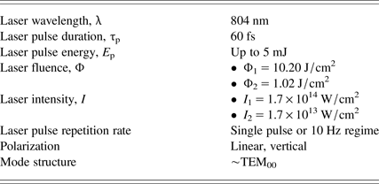
Laser irradiation experimental setup was similar to the one given in reference (Trtica et al., Reference Trtica, Batani, Redaelli, Limpouch, Kmetik, Ciganovic, Stasic, Gakovic and Momcilovic2013). Laser beam with diameter of 15 mm was focused normally on the target by a 150 mm focal length lens. During the irradiation process, laser was operated in the near fundamental transverse mode TEM00. Irradiations were carried out in vacuum (~0.001 mbar) environment. The employed Ti:Sapphire femtosecond laser system was PULSAR by Amplitude Technologies, based on chirped pulse amplification. Central laser wavelength is ~804 nm, maximum laser energy 12 mJ, and minimum laser spot diameter after focusing 50 µm (spot diameter in the experiments was typically ~250 µm, i.e. the target was positioned in front of the lens focus). The sample (ASP 30 steel) was mounted on a four axis (x, y, z, θ) motorized computer-controlled positioning system, so that the fresh target surface could be exposed to laser beam before each series of shots. Typical laser output parameters with experimental conditions are presented in Table 1.
Typical parameters of the femtosecond Ti:sapphire laser used for irradiation of the sample (ASP 30 steel)

Various analytical techniques were used for characterization of the sample before and after laser irradiation. Surface morphology was monitored by optical and scanning electron microscope (SEM), which was used in the secondary and back scattered electron modes. SEM was connected to an energy dispersive analyzer (EDS) for surface compositional analysis of the target. Also, the chemical composition of the target surface was determined by an Inductively Coupled Plasma-Optical Emission Spectrometer (ICP-OES). Interferometric profilometry was employed for specifying the geometry of the ablated area.
3. RESULTS AND DISCUSSION
Non-irradiated surface of the ASP 30 steel target, typically black-gray colored, had roughness of <0.02 µm, as obtained by profilometer. Elemental analysis of the surface, performed by EDS before irradiation showed the following content: Mo (~5.0 wt.%), Cr (~4.4 wt.%), W (~6.7 wt.%), V (~2.8 wt.%), Co (~8.4 wt.%), C (~4.7 wt.%), and iron balanced to 100 wt.%. Complete elemental analysis was normalized. It should be emphasized that ICP-OES analysis confirmed the target chemical content obtained with EDS method and both of them verify the elemental sample content given by the producer.
Generally, surface modification of the target depends on the laser output parameters – pulse energy density (fluence), intensity, wavelength, laser pulse duration, number of accumulated laser pulses, as well as the sample characteristics, for example absorptivity, and environmental conditions such as working atmosphere (vacuum, air, etc). Morphological changes of the ASP 30 steel target in vacuum atmosphere at the wavelength of 804 nm and as a function of accumulated laser pulses (1, 5, 10, 50, and 100 pulses) are shown in Figures 1–5. Figures 1, 2 and 4, 5 represent the modified surface of the sample at higher and reduced laser fluence/intensity, respectively. The induced features are presented in more details below.
SEM analysis of the ASP 30 steel surface after irradiation with fs laser pulses. Vacuum atmosphere; I 1 = 1.7 × 10¹⁴ W/cm2 (Φ1 = 10.20 J/cm2). A – view of the target before irradiation. B, C, D, E, – after irradiation with 1, 5, 10, and 100 pulses. (B1, C1, D1, E1 – entire spot; B2, C2, D2, E2 – central area and B3, C3, D3, E3 – periphery).

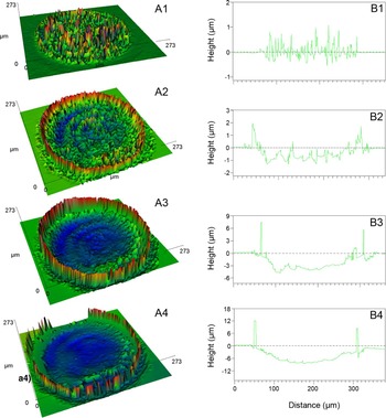
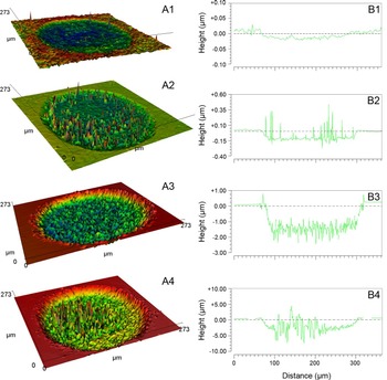
Profilometric analysis. 3D-(A) and 2D-view (B) of the damages and their cross section after laser irradiation of ASP 30 steel. Vacuum atmosphere; I 1 = 1.7 × 10¹⁴ W/cm2 (Φ1 = 10.20 J/cm2). (A1) and (B1), (A2) and (B2), (A3) and (B3), (A4) and (B4) – after irradiation with 1, 10, 50, and 100 pulses.

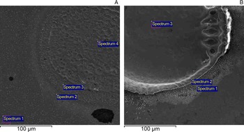
SEM images of the ASP 30 steel target with labeled locations for EDS analysis. I 1 = 1.7 × 10¹⁴ W/cm2 (Φ1 = 10.20 J/cm2). (A) Spectrum 1- non-irradiated region; after 1 pulse (spectra 2 and 3 – damage rim, that is, further and near periphery; spectrum 4 – damage center); (B) after 100 pulses (spectra 1, 2 – periphery region and spectrum 3 – central damage zone).

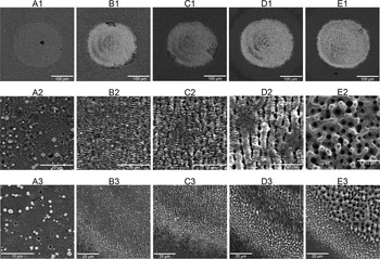
SEM analysis of the ASP 30 steel surface after irradiation with fs laser pulses. Vacuum atmosphere; I 2 = 1.7 × 10¹³ W/cm2 (Φ2 = 1.02 J/cm2). A, B, C, D, E, – after irradiation with 1, 5, 10, 50, and 100 pulses. (A1, B1, C1, D1, E1 – entire spot; A2, B2, C2, D2, E2 – central area and A3, B3, C3, D3, E3 – periphery).

Profilometric analysis. 3D-(A) and 2D-view (B) of the damages and their cross section after laser irradiation of ASP 30 steel. Vacuum atmosphere; I 1 = 1.7 × 10¹³ W/cm2 (Φ1 = 1.02 J/cm2). (A1) and (B1), (A2) and (B2), (A3) and (B3), and (A4) and (B4) – after irradiation with 1, 10, 50, and 100 pulses.

3.1. Regime of higher laser intensity
Initially, the effects of femtosecond laser on the sample were studied at higher laser intensity. This intensity was achieved on the sample surface by slightly focused laser beam with single pulse energy ~5 mJ and the corresponding fluence (Φ1) ~10.20 J/cm2. Equivalent laser intensity I 1 was ~1.7 × 10¹⁴ W/cm2 (Table 1). The view of the ASP 30 steel surface before and after irradiation is shown in Figure 1. Surface changes can be summarized as follows: (a) Action of 1, 5, 10, 50-not shown, and 100 pulses (Fig. 1B–1E) clearly resulted in morphological alterations; (b) even irradiation with 1 laser pulse, Figure 1B, caused drastic surface variations: (i) Evolution of lifted material in the central zone as well as formation of mini-craters within, (ii) production of needle like forms implying the possible formation of iron martensite structure (Trtica et al., Reference Trtica, Gakovic, Nenadovic and Mitrovic2001), (iii) appearance of hydrodynamic effects and, (iv) generation of the ripples (peripheral area), that is laser induced periodic surface structures (LIPSS). The spacing between two successive ripples (periodicity) was ~0.83 µm (Fig. 1B3), almost in accordance with the laser wavelength used (804 nm); (c) increased pulse count (Fig. 1C–1E) resulted in more prominent changes such as appearance of craters and barriers at central and peripheral zone, respectively. Clear presence of barriers at the rim can be attributed to intensive redeposition of ablated material.
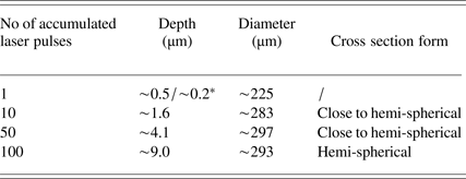
Depth and diameter of the damages, Table 2 and Figure 2, increased up to 100 accumulated laser pulses. It should be mentioned that the presence of some ablated material in the interaction region is recorded typically for lower pulse count (e.g., one pulse). The highest depth of ~9.0 µm (after 100 pulses, Fig. 2A4, B4) was achieved in the central part of the damage corresponding to the spatial profile of laser beam, that is TEM00 operation mode. Cross section of the damages, Table 2 and Figure 2, in one approximation is the same – it has hemi-spherical form (Fig. 2B2–B4). Similar form of the crater cross section on laser irradiated materials was also observed in reference (Yoo et al., Reference Yoo, Jeong, Greif and Ruso2000).
Damage parameters (depth, diameter and cross section) in a function of the laser pulse count

* ~0.5 and ~0.2 µm represents the maximal and minimal level of the materials, respectively (see Fig. 2B1).
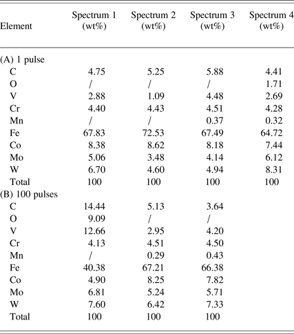
Apart from morphological features, chemical surface changes of ASP 30 steel (after irradiation by laser; vacuum ambience, ~0.001 mbar) were analyzed. These data at this high laser fluence and intensity (Φ1 = 10.20 J/cm2; I 1 = 1.7 × 10¹⁴ W/cm2) are insufficiently known in literature. EDS surface elemental analysis obtained from irradiated as well as non-irradiated locations is given in Table 3 and Figure 3. Mainly, the chemical surface changes on ASP 30 steel can be summarized as follows (Table 3): (i) Central zone (spectrum 4 and 3 – Fig. 3A, 3B, respectively) – reduction of Fe, C, and Co content compared with non-irradiated regions with simultaneous enhancement of Mo and W content. It is consistent with the assumption that during laser treatment of the surface, elements with lower boiling/melting temperature points (B/MTP) such as Fe, Co will be more efficiently removed than those with higher B/MTP like Mo, W; (ii) periphery zone (spectra 2, 3 and 1, 2 – Fig. 3A, 3B, respectively) – all elements which are included in ASP 30 steel were registered. After 100 pulses the barrier at rim region is built up (Fig. 2A4) and it is mainly composed of Fe, Co, Mo, and W (spectrum 2, Fig. 3B; Table 3), which is almost the same as in the nonirradiated sample implying that material from central area, during interaction, is redeposited in the barrier.
Local EDS elemental analysis of the irradiated and non-irradiated locations

Measuring locations are given in Fig. 3. (A) – spectrum 1- non-irradiated region. Spectra 2, 3 and 4 irradiated region after one laser pulse (spectra 2, 3-damage rim; spectrum 4-damage center); (B) – 100 laser pulses (spectra 1, 2-periphery region and spectrum 3-central damage zone).
Particularly, during laser–material interaction, the initial step of ablation comprises the absorption of laser radiation by free electrons (Bauerle, Reference Bauerle1996; Chichkov et al., Reference Chichkov, Momma, Nolte, Von Alvensleben and Tunnermann1996; Jeschke et al., Reference Jeschke, Garcia and Bennemann1999). Duration of this process is of the order of several tens of femtoseconds (Ahmmed et al., Reference Ahmmed, Grambow and Kietzig2014). Due to electron thermal conduction and diffusion, energy transfers to the lattice and the heat is transported into the material. Following the thermalization of the electron subsystem the energy transfers to electron-lattice subsystem with thermalization of this, latter subsystem at time scale of several hundred femtoseconds to several ps. Ablation of the material occurs at a time scale of tens of picoseconds after termination of the laser pulse.
If the laser intensity in front of the material is high, as in our case, the plasma can be created at very high temperature. This plasma can sometimes cause the so-called shielding effect, since the plasma itself absorbs large part of the incoming laser radiation. This effect, however, is characteristic for laser pulses in nano- and picosecond time domain, while for fs pulses it is not present, or drastically reduced, since the pulse ends before the plasma can be formed. The pulse energy is, therefore, deposited in the material without any substantial participation of laser–plasma interaction (Semerok et al., Reference Semerok, Salle, Wagner and Petite2002).
Additionally, during fs laser–target interaction, it should be taken into account that three operational regimes, depending on the laser intensity used, can be distinguished – low (I ~ 1010 W/cm2), high (I ~ 1015 W/cm2), and ultra-high laser intensity regime (I ~ 1019 W/cm2) (Torrisi, Reference Torrisi2011). The fs laser–steel interaction, in our case, is close to the second operational regime. In all of the stated regimes, the target is ablated while the fractional ionization is relatively different, that is low in the first one, and about 95 and 100% in the second and third regime (Torrisi, Reference Torrisi2011). Also, the ion energy, temperature, and electron densities are different so, in the first regime, these values are 100 eV/charge state 100 eV, and 1016/cm3, in the second 100 keV/charge state, 10 keV and 1020/cm3, and in the third 1–10 MeV/charge state, 10 keV and 1022/cm3 (Torrisi, Reference Torrisi2011). Also, it should be pointed out that, in our case, ablation process probably includes one additional mechanism aside the thermal (Gamaly et al., Reference Gamaly, Rode, Luther-Davies and Tikhonchuk2002). When an intensive field of laser acts on the material, it causes significant electron photo-emission from the surface. The surface is no longer quasineutral, which causes generation of a strong quasi-static electric field. When this field exceeds the binding energy of atoms and molecules, the material is disintegrated and removed from the surface. This phenomenon is known as electrostatic ablation.
3.2. Regime of reduced laser intensity
Surface modification of ASP 30 steel was monitored at lower laser intensity as well. For these irradiations, laser energy was attenuated about 10 times by neutral density filters placed in the laser beam path before the focusing lens. The given intensity was achieved on the sample surface by focused laser beam with the single pulse energy of 0.5 mJ (fluence Φ2 = 1.02 J/cm2). Equivalent laser intensity I 2 was 1.7 × 10¹³ W/cm2 (Table 1). The view of the ASP 30 steel sample surface after irradiation, with reduced laser energy, is shown in Figure 4.
Surface variations and phenomena can be summarized as follows: (a) Appearance of the damage already after 1 pulse (Fig. 4A); (b) increased damage depth and appearance of periodic surface structures especially in the central zone, except for 100 laser pulses, Figure 4A2, B2, C2, D2; (c) the border between irradiated and non-irradiated zone is very sharp. The expelled material accumulation, rim area, is drastically reduced compared with higher laser intensity of I 1 = 1.7 × 10¹⁴ W/cm2; (d) creation of plasma in front of the sample not as intensive as in the case of higher fluence. SEM micrographs (Fig. 4) show that the damages obtained using this, lower intensity, are less prominent. Focusing only on the features inside the damage area, for 1–50 pulses range (Fig. 4A2–D2), one can see the changes in morphological forms compared with the non-irradiated surface. Starting from 1 to 50 laser pulses (Fig. 4A–4D) the LIPSS were always produced in central irradiated zone. In case of 5 and 10 pulses (Fig. 4B2–C2) seems that two kinds of LIPSS are present (normal to one another) – more expressed for 10 pulses (periodicities of horizontal and vertical ripples were 0.7 and 1.7 µm, respectively). The average periodicity at 50 pulses, Figure 4D2, was about 3 µm. Accumulation of 100 pulses (Fig. 4E) resulted in LIPSS disappearance but in the formation of “bird's eye” features. Similar features at a target of Au film on sapphire substrate were recorded in reference (Nakata et al., Reference Nakata, Hiromoto and Miyanaga2010) and they were explained via interference phenomena of femtosecond laser beams. Generally, LIPSS is a universal phenomenon, which appears on any material that absorbs laser radiation (Young et al., Reference Young, Sipe and Van Driel1984). Their creation depends on many factors, such as laser properties (fluence, polarization, and coherence), angle of incidence relative to the sample, etc. Some literature data (Tan & Venkatakrishnan, Reference Tan and Venkatakrishnan2006) indicate that the origin of LIPSS can be attributed to the interference of incident laser beam with the so-called surface waves, scattered off the imperfections on the material and running along its surface. Generally speaking, LIPSS formation theory is rather complex and is dealt with in detail in reference by Bauerle (Reference Bauerle1996).
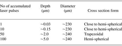
Depth, diameter, and cross-section evolution of the damages at the reduced laser energy/intensity is shown in Figure 5 and Table 4. Generally, the energy is deposited in the region with thickness of SD (skin depth) that is a function of the used laser intensity and fluence. In this context, the so called heat-affected zone, which is a maximum between SD and thermal diffusion depth (TDD), should also be mentioned (Kononenko et al., Reference Kononenko, Garnov, Pimenov, Konov, Romano, Borsos and Weber2000). SD is defined as the reciprocal value of absorption coefficient (α) while TDD = (D × τ)1/2, where D and τ represent the heat diffusivity and laser pulse duration, respectively. SD and TDD can be evaluated for our ASP 30 steel target. Using the value of 5.72 × 105 cm−1 (λ = 804 nm) for absorption coefficient of iron (Ordal et al., Reference Ordal, Bell, Alexander, Newquist and Querry1988), the calculated SD is ~17.5 nm for single pulse regime. TDD is of the order of ~0.65 nm based on D = 0.7 m2/s and fs pulse length. The D value is calculated based on the values for thermal conductivity, heat capacity, and density of ASP 30 steel. Larger SD than the TDD is expected due to the known fact that thermal effect in case of fs lasers is drastically reduced. Taking into account these calculations, it is possible to conclude that after first several laser pulses, for example 10 (and appropriate laser intensity), the ablation depth should be ~175 nm. Results presented in Figure 5 confirm this assumption.
Damage parameters (depth, diameter and cross section) in a function of the laser pulse count.

Based on the depth measurements, the damage threshold (DT), defined as the minimum laser radiation density/fluence necessary to induce detectable damage on the surface after one laser pulse, was evaluated to be around 0.20 J/cm2, which is in accordance with the literature data. For example, in case of femtosecond laser (150 fs) acting on stainless steel target, DT was determined to be 0.21 J/cm2 (Mannion et al., Reference Mannion, Magee, Coyne, O'Connor and Glynn2004). Maximum depth of the damage, Table 4, of 5 µm was achieved (after 100 laser pulses) while the cross section altered from the hemi-spherical to the trapezoidal form (Fig. 5). Profilometric analysis (Fig. 5), obtained at the regime of reduced laser energy/intensity, showed high-precise vertical and lateral modification/processing of the steel meaning that with further laser energy decrease and with a simultaneous choice of the number of accumulated pulses, extra-high precision can be achieved, imperative for some applications, for example mechanics (micro- and nanofabrication).
Chemical surface analysis of ASP 30 steel, after irradiation with lower laser energy/intensity, observed with SEM (back-scattered electron mode) showed no difference in elemental content between the irradiated and the non-irradiated surface. The irradiation of the sample in this intensity mode was also accompanied with appearance of low intensity plasma in front of the target.
4. CONCLUSIONS
The study of interaction of femtosecond laser with ASP 30 steel at 1014 and 1013 W/cm2 laser intensities, at varying number of laser pulses, is presented. Experiments were conducted in vacuum ambient. It was shown that both laser intensities induce specific morphological changes on the steel surface. Treatment at higher intensity of 1014 W/cm2 resulted in deep crater formation already after ten pulses. The reduced intensity of 1013 W/cm2, after ten pulses, led also to the creation of damage possessing superficial character and showing specific structure, that is laser induced periodic surface structures. The LIPSS, at reduced intensity, were created in pulse count range from 1 to 50. The presence of craters and LIPSS can have application in mechanical industry (fabrication of very precise apertures with small diameter) as well as micro-, nano-electronics, optics, etc. This study has shown that by careful choosing of the laser intensity and pulse count precise superficial material removal can be achieved. Thus, laser intensity ~1013 W/cm2 and low pulse count can lead to ultra-precise surface processing.
Chemical elemental analysis of the steel surface showed that changes are present only after irradiation with higher laser intensity of 1014 W/cm2. In the central irradiated zone, the content of elements with typically low boiling/melting temperature points (B/MTP), such as Fe and Co, was decreased, while the content of elements with higher B/MTP like Mo and W was increased, implying the possible chemical surface tailoring. The used laser intensities induced plasma in front of the steel target emitting in wide (including X-ray) spectral range.
Acknowledgments
The research was sponsored by: (i) The Ministry of Education, Science and Technology of the Republic Serbia through project “Effects of Laser Radiation on Novel Materials in Their Synthesis, Modification, and Analysis” (project No. 172019); (ii) COST- MP1208 action (“Developing the Physics and the Scientific Community for Inertial Confinement Fusion at the Time of NIF ignition”); (iii) Ministry of Education, Youth and Sports of the Czech Republic (projects LD14089 and LG15013).