




No CrossRef data available.
Article contents
Light microscopical structure and ultrastructure of a Besnoitia sp. in the naturally infected lizard Ameiva ameiva (Teiidae) from north Brazil, and in experimentally infected mice
Published online by Cambridge University Press: 28 November 2001
Abstract
A Besnoitia species of the teiid lizard Ameiva ameiva (L.), from north Brazil was established in laboratory mice and hamster by the intraperitoneal inoculation of bradyzoites in the tissue cysts. In the lizards all the cyst wall layers were closely apposed. In the mice the layers of the wall were distinguishable, and ultrastructurally the inner cytoplasmic layer contained either a tight network of endoplasmic reticulum or packed mitochondria or both. These components were less frequent or sparse in the inner cytoplasmic layer of cysts in the lizard. The only animals available for experiments in attempts to indicate the definitive host of the parasite were 3 kittens of the domestic cat and a juvenile specimen of the snake Boa constrictor raised in captivity. No evidence of infection could be detected in these animals after feeding them with the tissues of mice harbouring cysts with very large number of bradyzoites.
Keywords
Information
- Type
- Research Article
- Information
- Copyright
- © 2002 Cambridge University Press
INTRODUCTION
Tissue cysts of Besnoitia in saurian hosts have been described in light microscopical studies on a parasite named Besnoitia panamensis in the lizard Ameiva ameiva from Panama (Schneider, 1965, 1967a) and another referred to a B. saurina in the iguanid lizard Basiliscus basiliscus from Belize (Garnham, 1966). Based on the results of cross-immunity experiments in mice, Schneider (1967b) concluded that B. panamensis was a synonym of B. darlingi (Brumpt, 1913), described in a variety of mammals, and Levine (1988) regarded B. saurina as yet another synonym of this parasite. Schneider (1967a) showed that intraperitoneal inoculation of bradyzoites into mice resulted in proliferation of tachyzoites within cells of the peritoneal fluid, followed by the development of tissue cysts with which passage could be continued in further mice.
Ultrastructure of the proliferative forms of another Besnoitia species, B. jellisoni (Frenkel, 1953), has been studied in the peritoneal fluid cells of experimentally infected mice by Sheffield (1966). Gobel et al. (1985) described the fine structure of similar stages of B. besnoiti (Marotel, 1913) in cell culture. Available fine structural accounts of tissue cysts in natural infections are those concerning a Besnoitia sp., found in the eyelids of goats (Heydorn, et al. 1984) and another from the dermal and subdermal tissues of reindeer, Rangifer t. tarandus (Ayroud, Leighton & Tessaro, 1995). Fine structure studies of the cystic stages are available on the development of B. jellisoni in experimentally infected mice (Sheffield, 1968; Senaud, 1969). Tissue cysts of B. wallacei, B. besnoiti and B. darlingi were studied only by light microscopy by Frenkel (1977) and Bigalke (1981) and by Smith & Frenkel (1984), respectively. In the present communication we record the presence of Besnoitia tissue cysts in the teiid lizard, Ameiva ameiva, from north Brazil, and the successful establishment of the parasite in laboratory mice. The ultrastructure of the tissue cysts is described in the saurian and murine hosts, together with that of the proliferative stages in the peritoneal fluid cells of newly infected mice. Attempts have been made to determine the definitive host of the parasite.
MATERIALS AND METHODS
Adult specimens of Ameiva ameiva (L) were collected from open country in the vicinity of the town of Capanema, Para State, north Brazil for a general study of coccidian parasites. Tissue containing Besnoitia cysts found in necropsied lizards were fixed for histology and transmission electron microscopy (TEM).
Infection of mice
Tissue cysts were ground in 0.85% NaCl and approximately 0.1 ml of the suspension inoculated into mice by the intraperitoneal route. No attempt was made to calculate the number of bradyzoites inoculated. Some mice were sacrificed 8 days post-inoculation (p.i.) and peritoneal fluid smears prepared by pressing glass-slides lightly on the surface of the exposed viscera. The smears were rapidly air-dried and fixed for Giemsa's staining. Peritoneal fluid from infected mice at 8 days p.i. was centrifuged and the resulting pellet of cells was processed for TEM.
To study cyst production, other mice were killed at periods up to 34 days p.i. and pieces of the viscera and mesenteries were fixed for histology and electron microscopy.
Attempts to indicate a definitive, predator host
The tissues of chronically infected mice were fed to 3 newly weaned kittens and 1 young Boa c. constrictor, born in captivity. Whenever available, the faeces of these animals were checked for oocysts by direct examination and zinc sulphate flotation. One kitten was sacrificed 10 days later, and the other two 14 and 15 days later, respectively. A search for evidence of developmental stages or oocysts of Besnoitia was made by examining fresh scrapings of the intestinal epithelium along its length. The snake was killed and examined by the same method, 26 days after ingesting the infected mouse.
Processing of smears and tissues
Smears were rapidly air-dried and fixed either in absolute methyl alcohol for Giemsa's staining, or were fixed in aqueous Bouin's fluid for 20 min, washed in 70% ethyl alcohol until colourless and stained by a modified Giemsa's technique (Lainson, 1959).
For histology, tissues were fixed in neutral buffered formalin, dehydrated in ascending concentrations of ethyl alcohol and embedded in glycol–methacrylate medium (‘GMA' of Agar Co., UK). Histological sections, cut at a thickness of 2–3 μm with a glass knife on a Sorval JB4 microtome, were stained either with Meyer's haemalum–eosin (MH–E) or by Giemsa's stain after 20 min incubation in aqueous Bouin's fluid, followed by rinsing in water and 70% ethanol.
For TEM study materials were fixed in 2.5% glutaraldehyde in cacodylate buffer (0.1 M, pH 7.4) for 24 h at 4 °C, then rinsed repeatedly in the same buffer, post-fixed in 1.0% osmium tetroxide in the same buffer for 1 h and, after rinsing in the buffer, dehydrated in graded ethyl alcohol and embedded in Agar 100 medium (Agar Scientific Ltd, UK). Thin sections, cut on a Reichert ‘Ultracut’ microtome with a diamond knife, were stained on grids with uranyl acetate and lead citrate and examined with a Jeol 100CX TEM.
RESULTS
Only 2 of 17 specimens of A. ameiva had Besnoitia cysts. The cysts were mainly scattered in the mesenteries and along the surface of the intestine, some were also noted on the surface of the liver and spleen. Cysts were up to 0.3 mm in diameter, readily visible to the naked eye.
Light microscope and TEM structure of tissue-cysts in the connective tissues of the lizard
Fresh and stained preparations of the cyst contents revealed vast numbers of bradyzoites (Fig. 1). In histological sections, the cyst wall is seen to have been reduced to a narrow homogenous wall enclosing the closely packed bradyzoites.

Fig. 1. Besnoitia sp., in the teiid lizard Ameiva ameiva. Bradyzoites in a smear made from a ruptured tissue cyst. Bouin fixation/Giemsa staining.
By TEM the wall was shown to be comprised of an inner cytoplasmic (‘host cell’) layer and an external fibrous layer (‘cyst wall’ of Sheffield, 1968). The densely granulated cytoplasmic layer contained either a single multi-lobed nucleus, or several grossly hypertrophic nuclei (Fig. 2A). Its contents varied in thickness and density from cyst to cyst and contained variable proportions of rough and smooth endoplasmic reticulum (ER), cisternae, lipid vacuoles, and mitochondria (Fig. 2A and B): in some cysts, the rough ER aggregates into concentric formations (Fig. 2C). The plasma membrane on the PV boundary is protruded into very delicate tubule-like extensions which form a distinct layer on the surface of the PV wall. A few cytoplasmic protrusions penetrate, sometimes deeply, into the apposed external fibrous layer (Fig. 2A). This outer layer contained longitudinal and transverse filamentous strands within variable amounts of ground substance: in older cysts these microfibrils may be in disarray (Fig. 2C). The fibrous material in some cyst walls appears to be organized into 2 layers (Fig. 2A).

Fig. 2. (A–E) Fine structure of a tissue cyst from Ameiva ameiva. (A) Overall view of the cyst wall layers: the cytoplasmic (‘host-cell’) (C), its nuclei (N) and the PV-lining tubuli (T); extension of the cytoplasmic layer into the ‘cyst wall’ (pr) and ‘cyst wall’ layers (W). A gap in the cyst wall is marked with an arrow. (B) Details of the cytoplasmic (C) and the PV-lining tubuli layers (T): rough (rer) and smooth (er) endoplasmic reticulum, mitochondria (t), lipid vacuoles (L) and vesicles (v). (C) RER (arrow) concentrically arranged in the cytoplasmic layer (C), the cyst wall (W) seems to undergo shrinking (aging). (D) Dividing parasite with a primordium of daughter cell (arrow), a, apical complex, r. rhoptry. (E) Bradyzoite with elongate rhoptries (r) and ‘enigmatic bodies (arrows).
Within the PV most of the parasites are fully developed bradyzoites, with a few dividing by endodyogeny and containing primordia of daughter cells. The latter had a distinct conoid, a few rhoptries (not readily seen in section planes), while micronemes were lacking (Fig. 2D). The bradyzoites possessed a distinct conoid, elongate rhoptries and numerous micronemes which fill the entire pre-nuclear zone: they also contained the ‘enigmatic bodies’ as described by Senaud (1969), electron-dense globules and amylopectin granules (Fig. 2E).
Infection in laboratory mice and hamsters
From 7–14 days after intraperitoneal inoculation of the cyst contents, mice showed signs of sickness, with ruffled fur, diminished activity and increasing ascites. Some mice died with very abundant ascitic fluid containing variable numbers of intracellular and free parasites (tachyzoites) (Fig. 3A and B). Surviving mice recovered their normal appearance and activity but, in all those examined, possessed developing cysts in the tissues of their viscera and mesenteries. Hamsters were shown to develop similar infections. Some died from the acute infection.

Fig. 3. (A and B) Paired and quadruple sets of tachyzoites in the peritoneal exudate of a laboratory mouse, 7 days after the intraperitoneal inoculation of bradyzoites, Bouin/Giemsa staining.
TEM of tachyzoites in the peritoneal fluid cells of mice, 8 days p.i.
Host cells were predominantly neutrophils and monocytes, which contained single organisms (Fig. 4A) in a state of endodyogeny (Fig. 4B), or the products of such division (Fig. 4C). All stages possessed a bilayered pellicle, and were lodged in a parasitophorous vacuole (PV) bounded by a single unit membrane which was sometimes fragmented (Fig. 4A and B). Some tachyzoites were seen to be coated by a glycocalyx-like aggregate of a granular nature. The PV contained variable amounts of tubular whorls (Fig. 4A) mixed with a globular residue (Fig. 4B): sometimes it also contained lysosomal fragments. In some of the host cells the mitochondria and Golgi elements were seen to be concentrated at the cytoplasmic periphery of the PV. Where the PV boundary was indiscernible the outlining host cell cytoplasm also appeared to be disintegrating, with the material appearing to become continuous with that of the PV (Fig. 4C).

Fig. 4. (A–D) TEM of tachyzoites in the peritoneal fluid cells (monocytes) of mice, 8 days p.i. (A) Single tachyzoite in a host cell (H), the PV wall is discontinuous (arrows), and sometimes aligned by ER(er); PV contains tubular whorls (tw). (B) Tachyzoite dividing by endodyogeny (ga, granular aggregates; Ht, host-cell mitochondrium; m, micronemes, pp, progeny primordia; r, rhoptries; t, parasite mitochondrium). (C) Progeny of endodyogeny (a, apical complex; ga, granular aggregates; r, rhoptries). (D) Progeny of polyendyogeny.
The conoid of the tachyzoites was accompanied by a circle of elongate rhoptries, which extended beyond mid-length of the entire organism (Fig. 4C). Micronemes were seen in the single and the dividing tachyzoites (Fig. 4A and B) but seemed to be very few in their offspring. A layer of electron-dense substance was accumulated in the most anterior part of the subpellicular zone (Fig. 4C). Eight microtubules could be traced in longitudinal sections of the tachyzoites. All of the organisms, and their progeny, contained either 1 large mitochondrion or several smaller ones. Those in various stages of endodyogeny contained either rudimentary offspring or more advanced ones with an apical complex (Fig. 4B). Rounded, granular aggregates (ribosomes?), which are present only in the daughter cells (Fig. 4C), could already be traced in the endodyogeny primordia (Fig. 4B). An accumulation of 8 tachyzoites seen in a single PV (Fig. 4D) suggests the additional existence of merogonous (polyendogenous) division. Products of this division contained many rhoptries and micronemes.
Stages of tissue cysts in mice
In a light microscopical study of sectioned tissues of mice sacrificed (or dying) up to 25 days p.i., developing tissue cysts found in the lungs, stomach serosa and mesenteries (Fig. 5A and B) measured up to 100 μm in diameter, while in other mice killed at 30–34 days p.i. some cysts had reached approximately 130×100 μm (Fig. 5C). The cyst wall seen at first to be composed of an inner cytoplasmic zone with numerous hypertrophic and segmented nuclei, each with several small nucleoli, and a peripheral non-nucleated and more hyaline layer. The 2 layers were separated by a space containing many minute, dark granules; the PV was filled with divided stages (Fig. 5A). With further development the PV, packed with parasite progeny, increased in volume and both the cyst wall inner nucleate and the apposing outer layers became compressed (Fig. 5B). Further increase in the cyst volume resulted in compression of the wall till the inner details of the 2 layers become only vaguely distinguishable (Fig. 5C). There were no notable changes in the host's surrounding tissues.

Fig. 5. (A–D) Histological view of stages of tissue-cysts in mice. (A) Young cyst (25 days p.i.) enclosing PV filled with products of endodyogeny, the nucleate cytoplasmic layer (C) clearly separated by a gap filled with fine granules (arrow) from the ‘cyst wall’ (W); (B) On-growing cyst (30 days p.i.), with the cytoplasmic, nucleate layer (C) being compressed against the ‘cyst wall’ (W). (C) Further differentiated cyst (30 days p.i.), with its compressed wall components. (D) TEM view of walls of tissue cysts in experimentally infected mice (30 days p.i.) (C, cytoplasmic layer; H, host connective tissue; pr, protrusions of the cytoplasmic layer; PV, parasitophorous vacuole, rer, rough ER; t, mitochondria; W, cyst wall).
TEM of tissue cysts from a mouse 30 days p.i.
The tissue cysts were at different stages of differentiation but demonstrated the same configuration of a PV filled with the products of endodyogeny enclosed by an inner cytoplasmic wall (‘host cell’) containing several large, compact or multilobed nuclei and an apposed outer, fibrous wall of 1 or more, layers (‘cyst wall’) (Figs 5D and 6A, B). The inner cytoplasmic layer contained rough ER, either concentrically tightly packed (Figs 5D and 6A), or loose and disarrayed (Fig. 6B). The spaces between the ER contained variable quantities of mitochondria (Figs 5D and 6C). ER elements were absent between the nuclei and the PV boundary, and in the septum extending to divide the PV of some cysts into 2 compartments. The plasma membrane on the PV boundary, similar to that seen in cysts in the lizard host, formed a layer of very delicate tubule-like extensions lining the PV wall surface (Fig. 6B). The membraneous boundary of the inner cytoplasmic layer was seen to send both short and elongated protrusions into the adjoining outer fibrous layer: these protrusions seemingly contained extensions of the ER (Figs 5D and 6A). The outer, fibrous layers of the cyst wall contained fibrillar bundles, sometimes orientated perpendicularly, and a narrow gap containing a fine ground substance separating the main fibrous layer from an outer narrower one (Fig. 5D).

Fig. 6. (A–C) TEM view of tissue cysts in experimentally infected mice (30 days p.i.), pr, protrusions of the cytoplasmic layer scale. (A) Enlarged view of the cytoplasmic wall with concentric rough ER, arrow head, PV-lining tubuli layer. (B) Cytoplasmic wall with mitochondria (t) and scattered rough ER (arrow heads). (C) Dividing parasite (T) and bradyocytes (B) in the PV of the tissue cyst shown in Fig. 5D; r, rhoptries.
The PV of each cyst contained bradyzoites, as well as a few stages undergoing division by endodyogeny (Fig. 6C). Dividing stages had rhoptries but lacked micronemes. Some bradyzoites were showing the expanded posterior end, with pellicular thickenings (‘posterior ring’ of Sheffield, 1968) characteristic of the bradyzoites' site of detachment from the mother cell.
Attempts to indicate the definitive host
No oocysts characteristic of Besnoitia were detected in faeces from the 3 kittens 4–15 days after they were fed with tissue cysts of chronically infected mice, or in fresh preparations of scrapings along the length of the intestine of each animal on autopsy. Similarly, no evidence of an induced infection could be found in the young boa-constrictor.
DISCUSSION
The external fibrous capsule (‘cyst-wall’ of Sheffield, 1968, or the ‘secondary cyst-wall’ of Heydorn et al. 1984) has the same basic fine structure as described by Heydorn et al. (1984) in cysts in natural infections of a Besnoitia sp. of goats, also as described by Ayroud et al. (1995) in reindeer and in cysts of Besnoitia jellisoni in experimentally infected laboratory mice Sheffield, 1968; Senaud, 1969; Scholtyseck, Mehlhorn & Muller, 1974).
The fine structure of the cyst's inner multinucleated cytoplasm (‘host-cell’) in the naturally infected lizard and the experimentally infected mice varies considerably in texture and components. The host-cell layer of cysts in our experimentally infected mice was densely filled by ER components and mitochondria. Some TEM images of tissue cysts from mice with 3- to 8-week-old infections of B. jellisoni described by Sheffield (1968) and Senaud (1969) showed a similar increase in ER and mitochondria in the host-cell layer. In other images, these components seem to be already in disarray. The cyst wall in the lizards was reminiscent of that reported in the natural infection of Besnoitia in goats and reindeer (Heydorn et al. 1984; Ayroud et al. 1995). These cysts are evidently long lasting, in their host-cell layer the vesiculate, ER is disarrayed and the mitochondria are few in number.
A high aggregation of ER and mitochondria is suggestive of accelerated metabolic activity. Parasites appear to induce the host-cell to assist actively in their nutrition (Trager, 1974), and Marquardt, Bryan & Long (1984) suggested that coincidental nuclear and nucleolar enlargement in host-cells harbouring intestinal coccidia is linked with a high level of transcription. Cellular and nuclear hypertrophy of parasitized cells is characteristic of some other coccidial infections: Eimeria, for example (Fernando & Pasternak, 1977), and ‘globidia’ (Mehlhorn, Senaud & Heydorn, 1984; Senaud Heidorn & Mehlhorn, 1984; Paperna, 1999). On the other hand, it does not occur in infections with Toxoplasma or Hammondia (Dubey, 1977).
Dividing parasites were far more numerous in the young cysts (in mice), but the presence of some apparently fully developed bradyzoites suggests that young cysts are also infective. Only a few dividing parasites were seen among the bradyzoites of the older cysts in the lizards. The fine structure of both dividing forms and bradyzoites was similar to that previously described for Besnoitia by Shefield (1968), Senaud (1969), Scholtyseck, Mehlhorn & Muller (1968) and Heydorn et al. (1984). The polyendodygenous stages as seen by Senaud (1969) in B. jellisoni were not found in our material.
The fine structure of tachyzoites in the peritoneal exudate of mice infected with Besnoitia from A. ameiva closely resembled that described for B. jellisoni in the same animal, by Sheffield (1966). The tachyzoites are reminiscent of Toxoplasma gondii, which similarly develop in the peritoneal exudate cells of inoculated mice. Both parasites divide by endodyogeny, and both form ‘clone’ progenies of 8 tachyzoites sharing the same PV and suggesting a simultaneous division, or endopolygeny (Dubey & Beattie, 1988). A process of endopolygeny yielding rosettes, and concurrent with endodyogeny, has also been shown to occur in B. besnoiti growth in cultured cells (Gobel et al. 1985).
The specific identity of the Besnoitia sp., from the lizard A. ameiva in Amazonian Brazil remains questionable. Schneider (1965) gave the name of B. panamensis to the parasite from Ameiva in Panama but, based on the results of cross-immunity experiments in mice, he later concluded that this was synonymous with B. darlingi (Brumpt, 1913) of opossums which he found in the same geographical locality. Our detection of tissue-cysts of Besnoitia in Ameiva in the present study tempts one to regard the Panamanian and Brazilian parasites as conspecific. The range of different intermediate hosts for species of Besnoitia, however, is extensive (Levine, 1988) and it may well be that different species of this parasite may infect the same secondary host.
The definitive hosts of B. besnoiti, B. wallacei and B. darlingi are considered to be felids (Peteshev, Galuzo & Polomoshnov, 1974; Wallace & Frenkel, 1975; Smith & Frenkel, 1984) and the domestic cat has been used experimentally to study the life-cycle of these parasites. On the other hand, Diesing et al. (1988) reported failure to infect domestic cats, among a variety of other carnivorous mammals, snakes and vultures, fed with the tissue cysts of a parasite considered to be B. besnoiti from cattle. They concluded that none of these are definitive hosts and that the life-cycle of the parasite remains obscure. In addition, Ayroud et al. (1995) recorded no shedding of oocysts by 2 cats fed with the cysts of a Besnoitia species from reindeer in Canada.
In view of these authors' findings and our own failure to produce a detectable infection in 3 kittens fed with mice harbouring tissue cysts of the Besnoitia of Ameiva we are led to the conclusion that in this case, too, cats are not the definitive hosts.
Our failure to obtain evidence of development of the Besnoitia sp., in the single boa-constrictor does not exclude the possibility of another species of snake being the definitive host. A number of different species of snakes commonly eat lizards, and we have indicated a snake to be the definitive host of a Sarcocystis sp., commonly found in Ameiva (Lainson & Paperna, 2000).
This work was supported by a grant from the Wellcome Trust, London (to R.L.). We thank Constancia M. Franco and Manoel C. de Souza, Instituto Evandro Chagas for technical help in the field and laboratory and Marina Schein, Department of Animal Sciences, H.U.J. for histological and electron microscopical laboratory assistance.
References
AYROUD, M., LEIGHTON, F. A. & TESSARO, S. V. (1995). The morphology and pathology of Besnoitia sp. in reindeer (Rangifer tatrandus tarandus). Journal of Wildlife Diseases 31, 319–325.CrossRefGoogle Scholar
BIGALKE, A. (1981). Besnoitiosis and globidiosis. Current Topics in Veterinary Medicine and Animal Science 6, 429–442.Google Scholar
DIESING, L., HEYDORN, A. O., MATUSCHKA, F. R., BAUER, C., PIPANO, E., DE WAAL, D. T. & POTGIETER, F. T. (1988). Besnoitia besnoitii: studies on the definitive host and experimental infections in cattle. Parasitology Research 75, 114–117.CrossRefGoogle Scholar
DUBEY, J. P. (1977). Toxoplasma, Hammondia, Besnoitia, Sarcocystis, and other cyst-forming Coccidia of man and animalsParasitic Protozoa, Vol. III (ed. KREIER, J. P.), 101–237Academic PressNew York.
DUBEY, J. P. & BEATTIE, C. P. (1988). Toxoplasmosis of Animals and Man. CRC Press, Boca Raton, Florida.
FERNANDO, M. A. & PASTERNAK, J. (1977). Isolation of host-cell nuclei from chick intestinal cells infected with second-generation schizonts of Eimeria necatrix. Parasitology 74, 27–32.CrossRefGoogle Scholar
FRENKEL, J. K. (1977). Besnoitia wallacei of cats and rodents; with reclassification of other cyst-forming isosporoid coccidia. Journal of Parasitology 63, 611–628.CrossRefGoogle Scholar
GARNHAM, P. C. C. (1966). Besnoitia (Protozoa: Toxoplasmea) in lizards. Parasitology 56, 329–334.CrossRefGoogle Scholar
GOBEL, E., WIDAUER, R., REIMANN, M. & MUNZ, E. (1985). Ultrastructure of asexual multiplication of Besnoitia besnoiti (Marotel, 1912) in Vero- and CRFK-cell cultures. Zentrablatt fuer Veterinärmedizin B 32, 202–212.CrossRefGoogle Scholar
HEYDORN, A. O., SENAUD, J., MEHLHORN, H. & HEINONEN, R. (1984). Besnoitia sp. From goats in Kenya. Zeitschrift fuer Parasitenkunde 70, 709–713.Google Scholar
LAINSON, R. (1959). Atoxoplasma Garnham 1950, as a synonym for Lankesterella Labbe 1889. Its lifecycle in the English sparrow (Passer domesticus domesticus Linn). Journal of Protozoology 6, 360–371.Google Scholar
LAINSON, R. & PAPERNA, I. (2000). The life cycle and ultrastructure of Sarcocystis ameivamastigodryasi n. sp., in the lizard Ameiva ameiva (Teiidae) and the snake Mastigodryas bifossatus (Colubridae). Parasite 7, 263–274.Google Scholar
LEVINE, N. D. (1988). The Protozoan Phylum Apicomplexa, Vol. II. CRC Press, Boca Raton, Florida, USA.
MARQUARDT, W. C., BRYAN, J. H. D. & LONG, P. L. (1984). Nucleolar hypertrophy as an indicator of transcription in cells infected with second generation meronts of Eimeria tenella. Journal of Protozoology 31, 569–574.CrossRefGoogle Scholar
MEHLHORN, H., SENAUD, J. & HEYDORN, A. O. (1984). Two types of globidium cysts of goats. Zeitschrift fuer Parasitenkunde 70, 731–737.CrossRefGoogle Scholar
PAPERNA, I. (1999). Globidia infection in the gut of an Australian gecko Heteronotia binoei. Folia Parasitologica 46, 175–178.Google Scholar
PETESHEV, V. M., GALUZO, I. G. & POLOMOSHNOV, A. P. (1974). Cats – definitive hosts of Besnoitia (Besnoitia besnoiti) (in Russian). Izvestiae Akademii Nauk KazakhestanSSR, B 1, 33–38.Google Scholar
SCHNEIDER, C. R. (1965). Besnoitia panamensis, sp. n. (Protozoa: Toxoplasmatidae) from Panamian lizards. Journal of Parasitology 51, 340–344.Google Scholar
SCHNEIDER, C. R. (1967a). The distribution of lizard besnoitiosis in Panama, and its transfer to mice. Journal of Protozoology 14, 674–678.Google Scholar
SCHNEIDER, C. R. (1967b). Cross-immunity evidence of the identity of Besnoitia panamensis from lizards and B. darlingi from opossums. Journal of Parasitology 53, 886.Google Scholar
SCHOLTYSECK, E., MEHLHORN, H. & MULLER, B. E. G. (1968). Identifikation von Merozoites der vier cystenbilden Coccidien (Sarcocystis, Toxoplasma, Besnoitia, Frenkelia) auf Grund feinstructureller Kriterien. Zeitschrift fuer Parasitenkunde 42, 185–206.Google Scholar
SCHOLTYSECK, E., MEHLHORN, H. & MULLER, B. E. G. (1974). Feinstructur der Cyste und Cystenwand von Sarcocystis tenella, Besnoitia jellisoni, Frenkelia sp. und Toxoplasma gondii. Journal of Protozoology 21, 284–294.CrossRefGoogle Scholar
SENAUD, J. (1969). Ultrastructure des formations kystiques de Besnoitia jellisoni (Frenkel, 1953) protozoaire, Toxoplasmea, parasite de la suris (Mus musculus). Protistology 5, 413–430.Google Scholar
SENAUD, J., HEYDORN, A. O. & MEHLHORN, H. (1984). Three types of globidium of sheep: an in vivo and in vitro investigation. Zeitschrift fuer Parasitenkunde 70, 721–729.CrossRefGoogle Scholar
SHEFFIELD, H. G. (1966). Electron microscopic study of the proliferative form of Besnoitia jellisoni. Journal of Parasitology 52, 583–594.CrossRefGoogle Scholar
SHEFFIELD, H. G. (1968). Observation on the fine structure of the ‘cyst stage’ of Besnoitia jellisoni. Journal of Protozoology 15, 685–693.CrossRefGoogle Scholar
TRAGER, W. (1974). Some aspects of intracellular parasitism. Science 183, 269–283.CrossRefGoogle Scholar
SMITH, D. D. & FRENKEL, J. K. (1984). Besnoitia darlingi (Apicomplexa, Sarcocystidae, Toxoplasminae): Transmission between opossum and cat. Journal of Protozoology 31, 584–587.CrossRefGoogle Scholar
WALLACE, G. D. & FRENKEL, J. K. (1975). Besnoitia species (Protozoa, Sporozoa, Toxoplasmatidae): recognition of cyclic transmission by cats. Science 188, 369–371.CrossRefGoogle Scholar

Fig. 1. Besnoitia sp., in the teiid lizard Ameiva ameiva. Bradyzoites in a smear made from a ruptured tissue cyst. Bouin fixation/Giemsa staining.
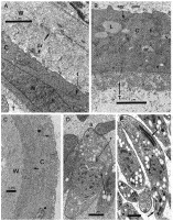
Fig. 2. (A–E) Fine structure of a tissue cyst from Ameiva ameiva. (A) Overall view of the cyst wall layers: the cytoplasmic (‘host-cell’) (C), its nuclei (N) and the PV-lining tubuli (T); extension of the cytoplasmic layer into the ‘cyst wall’ (pr) and ‘cyst wall’ layers (W). A gap in the cyst wall is marked with an arrow. (B) Details of the cytoplasmic (C) and the PV-lining tubuli layers (T): rough (rer) and smooth (er) endoplasmic reticulum, mitochondria (t), lipid vacuoles (L) and vesicles (v). (C) RER (arrow) concentrically arranged in the cytoplasmic layer (C), the cyst wall (W) seems to undergo shrinking (aging). (D) Dividing parasite with a primordium of daughter cell (arrow), a, apical complex, r. rhoptry. (E) Bradyzoite with elongate rhoptries (r) and ‘enigmatic bodies (arrows).
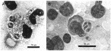
Fig. 3. (A and B) Paired and quadruple sets of tachyzoites in the peritoneal exudate of a laboratory mouse, 7 days after the intraperitoneal inoculation of bradyzoites, Bouin/Giemsa staining.
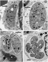
Fig. 4. (A–D) TEM of tachyzoites in the peritoneal fluid cells (monocytes) of mice, 8 days p.i. (A) Single tachyzoite in a host cell (H), the PV wall is discontinuous (arrows), and sometimes aligned by ER(er); PV contains tubular whorls (tw). (B) Tachyzoite dividing by endodyogeny (ga, granular aggregates; Ht, host-cell mitochondrium; m, micronemes, pp, progeny primordia; r, rhoptries; t, parasite mitochondrium). (C) Progeny of endodyogeny (a, apical complex; ga, granular aggregates; r, rhoptries). (D) Progeny of polyendyogeny.
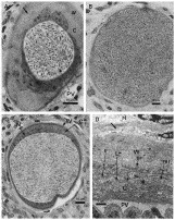
Fig. 5. (A–D) Histological view of stages of tissue-cysts in mice. (A) Young cyst (25 days p.i.) enclosing PV filled with products of endodyogeny, the nucleate cytoplasmic layer (C) clearly separated by a gap filled with fine granules (arrow) from the ‘cyst wall’ (W); (B) On-growing cyst (30 days p.i.), with the cytoplasmic, nucleate layer (C) being compressed against the ‘cyst wall’ (W). (C) Further differentiated cyst (30 days p.i.), with its compressed wall components. (D) TEM view of walls of tissue cysts in experimentally infected mice (30 days p.i.) (C, cytoplasmic layer; H, host connective tissue; pr, protrusions of the cytoplasmic layer; PV, parasitophorous vacuole, rer, rough ER; t, mitochondria; W, cyst wall).
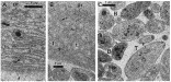
Fig. 6. (A–C) TEM view of tissue cysts in experimentally infected mice (30 days p.i.), pr, protrusions of the cytoplasmic layer scale. (A) Enlarged view of the cytoplasmic wall with concentric rough ER, arrow head, PV-lining tubuli layer. (B) Cytoplasmic wall with mitochondria (t) and scattered rough ER (arrow heads). (C) Dividing parasite (T) and bradyocytes (B) in the PV of the tissue cyst shown in Fig. 5D; r, rhoptries.
 You have
Access
- 9
- Cited by