Introduction
Patients with hoarding disorder (HD) show difficulty in discarding possessions and a tendency to accumulate a large number of objects, regardless of their real value, cluttering living areas (Timpano et al., Reference Timpano, Rasmussen, Exner, Rief, Schmidt and Wilhelm2013). Hoarding symptoms were first considered as diagnostic criteria for obsessive-compulsive personality disorder or a symptom dimension of obsessive-compulsive disorder (OCD). However, most individuals with OCD do not report significant hoarding behavior (Pertusa et al., Reference Pertusa, Frost and Mataix-Cols2010), and individuals with HD typically do not meet other symptom criteria for OCD (Frost et al., Reference Frost, Steketee and Tolin2012). Indeed, hoarding and OCD symptoms show weak correlations, and in factor analyses, they are typically categorized in separate dimensions (Wu and Watson, Reference Wu and Watson2005). Compulsive hoarding was therefore considered as an independent diagnosis, within the OCD spectrum, in the fifth edition of the Diagnostic and Statistical Manual of Mental Disorders (DSM-V) (APA, 2013).
The physiopathology of HD, however, is largely unknown. Most studies assessing the neural correlates of compulsive hoarding have evaluated hoarding symptoms (from a dimensional perspective) in individuals with OCD (Mataix-Cols et al., Reference Mataix-Cols, Wooderson, Lawrence, Brammer, Speckens and Phillips2004; Harrison et al., Reference Harrison, Pujol, Cardoner, Deus, Alonso, López-Solà, Contreras-Rodríguez, Real, Segalàs, Blanco-Hinojo, Menchon and Soriano-Mas2013), or have compared OCD samples with and without compulsive hoarding (Saxena et al., Reference Saxena, Brody, Maidment, Smith, Zohrabi, Katz, Baker and Baxter2004; An et al., Reference An, Mataix-Cols, Lawrence, Wooderson, Giampietro, Speckens, Brammer and Phillips2009). Therefore, they are not representative of HD patients not exhibiting obsessive-compulsive symptoms. Only more recent studies have compared individuals with hoarding without OCD to healthy controls (HCs) or OCD patients without hoarding symptoms (Tolin et al., Reference Tolin, Kiehl, Worhunsky, Book and Maltby2009, Reference Tolin, Stevens, Villavicencio, Norberg, Calhoun, Frost, Steketee, Rauch and Pearlson2012). However, these studies used tasks designed to trigger complex emotions during hoarding-related decision-making (i.e. discarding objects). Consequently, they failed to provide a meaningful comparison with OCD because of an overall lack of activation in these patients (Tolin et al., Reference Tolin, Stevens, Villavicencio, Norberg, Calhoun, Frost, Steketee, Rauch and Pearlson2012). To substantiate the clinical division between HD and OCD in neurobiological terms, it is important to compare the behavioral and brain activation features of both groups of patients during the performance of tasks relevant for OCD pathophysiology.
Poor cognitive control plays an important role in pathophysiological models of OCD and has been considered as a potential endophenotype of the disorder (Chamberlain and Menzies, Reference Chamberlain and Menzies2009). Impairments in inhibitory function and attentional switching could indeed underlie the poor control over obsessions and compulsions (Snyder et al., Reference Snyder, Kaiser, Warren, Heller, Hall and Hospital2015). These executive functions are known to be supported by prefrontal, parietal and striatal regions (Norman et al., Reference Norman, Carlisi, Lukito, Hart, Mataix-Cols, Radua and Rubia2016), which are central in prevailing neurobiological models of OCD as part of the cortico-striatal-thalamo-cortical circuitry (Menzies et al., Reference Menzies, Chamberlain, Laird, Thelen, Sahakian and Bullmore2008; van den Heuvel et al., Reference van den Heuvel, van Wingen, Soriano-Mas, Alonso, Chamberlain, Nakamae, Denys, Goudriaan and Veltman2016). Moreover, OCD is also characterized by excessive performance monitoring, which might underlie the emergence of particular OCD symptoms (e.g. repetitive checking) (Harkin et al., Reference Harkin, Miellet and Kessler2012). Performance monitoring has been linked to dorsolateral prefrontal and anterior cingulate cortices (dlPFC and ACC) (Melcher et al., Reference Melcher, Falkai and Gruber2008), and neuroimaging studies have consistently reported ACC hyperactivation in OCD during performance monitoring (Melcher et al., Reference Melcher, Falkai and Gruber2008).
Protocols evaluating these neurocognitive domains could therefore shed light on further discriminating HD from OCD. Nevertheless, only two previous neuroimaging studies have focused on the comparison of the neural correlates of executive dysfunction between OCD and HD. The first study assessed response inhibition and performance monitoring in a Go/No-Go protocol (Tolin et al., Reference Tolin, Witt and Stevens2014), while the second examined these same functions and also included a response conflict task (i.e. Stroop) (Hough et al., Reference Hough, Luks, Lai, Vigil, Guillory, Nongpiur, Fekri, Kupferman, Mathalon and Mathews2016). Neither of them found performance differences between groups. At the neurobiological level, HD patients showed significant hyperactivations in comparison with the OCD group during response inhibition, although the specific pattern of findings differed between studies: from a single cluster in the right precentral gyrus (Tolin et al., Reference Tolin, Witt and Stevens2014), to a more extended pattern encompassing the dlPFC, insula, visual cortex and cerebellum (Hough et al., Reference Hough, Luks, Lai, Vigil, Guillory, Nongpiur, Fekri, Kupferman, Mathalon and Mathews2016). During response conflict, HD participants exhibited hypoactivation of the medial PFC (Hough et al., Reference Hough, Luks, Lai, Vigil, Guillory, Nongpiur, Fekri, Kupferman, Mathalon and Mathews2016). Finally, during performance monitoring and error processing, differences were only observed in response inhibition, and they also diverged between studies: individuals with HD showed a hypoactivation of the orbitofrontal cortex (Tolin et al., Reference Tolin, Witt and Stevens2014) or a hyperactivation of the right vlPFC and the bilateral striatum (Hough et al., Reference Hough, Luks, Lai, Vigil, Guillory, Nongpiur, Fekri, Kupferman, Mathalon and Mathews2016).
Overall interpretation of above findings, involving different executive domains (i.e. response inhibition, response conflict and error processing), is unavoidably hampered by the use of different protocols, which could be a source of non-specific task-related heterogeneity. This is especially important considering that successful managing of response conflict also depends on response inhibition capacity. In the current study, to overcome task-related heterogeneity and better characterize each of the above executive domains, we assessed response inhibition, conflict and error processing with two variants of a cognitive control protocol (i.e. stop-signal and switch-signal) that were comparable in terms of performance demands and assessment of error processing. On the basis of previous research (Tolin et al., Reference Tolin, Witt and Stevens2014; Hough et al., Reference Hough, Luks, Lai, Vigil, Guillory, Nongpiur, Fekri, Kupferman, Mathalon and Mathews2016), we hypothesized that HD and OCD patients would show different patterns of brain activation across all domains, mostly in the frontal lobe. Nonetheless, since significant findings were observed in other brain areas as well, we also explored whole-brain patterns of activation to comprehensively characterize between-group differences. A HC group was also studied to provide reference comparison values.
Methods
Participants
Seventeen patients with HD (DSM-V criteria), 18 patients with OCD were recruited through the Departments of Psychiatry of Bellvitge University Hospital and Parc de Salut Mar (Barcelona, Spain). Patients were evaluated by a senior psychiatrist with extensive clinical experience in OCD and related disorders. Exclusion criteria for patients included being under the age of 18 or older than 65, the presence of a current psychotic disorder, a recent history (i.e. 6 months) of psychoactive substance abuse or dependence, personality disorders, mental retardation, any severe organic or neurological pathology except for tic disorder, and the presence of any contraindication to magnetic resonance imaging (MRI) scanning. Comorbidity with other disorders (i.e. mood or anxiety disorders) was not considered an exclusion criterion provided that either HD or OCD was the main reason for seeking medical assistance. All patients remained on stable medication doses for at least 3 months before the MRI acquisition. In addition, nineteen HCs (without a current or past history of a psychiatric disorder), of comparable age and gender, were recruited from the same centers according to the same exclusion criteria.
The Structured Clinical Interview for DSM-IV Axis I Disorders-Clinician Version (SCID-CV) (First et al., Reference First, Spitzer, Gibbon and Williams1996) was used to confirm OCD diagnosis (OCD group), to assess for comorbid disorders (clinical groups) and to discard mental health diagnoses (HCs). In the OCD group, the severity of obsessive-compulsive symptoms was assessed with the Spanish version of the Yale-Brown Obsessive-Compulsive Scale (YBOCS) (Goodman et al., Reference Goodman, Price, Rasmussen, Mazure, Fleischmann and Hill1989; Sal y Rosas et al., Reference Sal y Rosas, Vega-Dienstmaier, Mazzotti, Vidal, Guimas, Adrianzén and Vivar2002). HD diagnosis was confirmed using the Structured Interview for Hoarding Disorder (SIHD) (Nordsletten et al., Reference Nordsletten, de la Cruz, Pertusa, Reichenberg, Hatch and Mataix-Cols2013), and symptom severity was quantified using the validated Spanish versions of the Saving Inventory-Revised (SI-R) (Frost et al., Reference Frost, Steketee and Grisham2004; Tortella-Feliu et al., Reference Tortella-Feliu, Fullana, Caseras, Andión, Torrubia and Mataix-Cols2006) and the Hoarding Rating Scale-Self Report (HRS-SR) (Tolin et al., Reference Tolin, Frost, Steketee and Fitch2008). Moreover, all participants completed the revised Obsessive-Compulsive Inventory (OCI-R) (Foa et al., Reference Foa, Huppert, Leiberg, Langner, Kichic, Hajcak and Salkovskis2002; Fullana et al., Reference Fullana, Tortella-Feliu, Caseras, Andión, Torrubia and Mataix-Cols2005). Written informed consent was obtained from all participants after providing a complete description of the study, which was performed in accordance with the Declaration of Helsinki and approved by the local ethical review board in clinical research of Bellvitge University Hospital.
Task design
The study tasks are described in online Supplementary Fig. S1. A rapid, mixed trial, randomized presentation, event-related fMRI design was used for both the stop-signal and the switch-signal tasks, which were adapted from McClure et al. (Reference McClure, Treland, Snow, Schmajuk, Dickstein, Towbin, Charney, Pine and Leibenluft2005). The tasks were programed in E-prime (https://pstnet.com/products/e-prime) and visually presented through a laptop computer connected to MRI-compatible high-resolution goggles (VisuaStim Digital System, Resonance Technology Inc., Northridge, CA, USA). Subject responses were registered by means of right- and left-response grips based on optical fiber transmission (NordicNeuroLab Inc., Bergen, Norway). Both tasks had the same duration (9 min), which were divided into three 3-min sessions to avoid fatigue. Participants were instructed outside the scanner on how to complete the task and trained again inside the scanner immediately before beginning the task. Both tasks shared identical go trials that started with the presentation of a black screen for 750 ms followed by a white point that lasted 500 ms. Afterward, a figure appeared on the screen for 1000 ms and subjects had to confirm whether it was a circle, by pressing the left button, or a square, by pressing right button. Therefore, circles and squares were the go signals, which had a mean inter-stimulus interval of 1250 ms. Subjects were instructed to make their responses as soon as possible after stimulus presentation.
In the stop-signal task, motor response had to be selectively inhibited in the randomly presented stop trials in which figure presentation was interrupted at a mean time of 300 ms by a stop signal that lasted 700 ms. Importantly, throughout the task, a tracking algorithm increased or decreased (in 50 ms steps) the time interval between go- and stop-signals according to the performance of each subject in preceding trials to minimize individual differences in task difficulty. During the switch-signal task, subjects had to switch from the standard stimulus-response association (i.e. circle – left hand; square – right hand) to the opposite. That is, when a circle was followed by a switch signal, the subject had to use the right button instead of the left, and vice versa when a square was presented.
Each task consisted of 258 trials, 86 per session, with a duration of 2250 ms each. Sixty of those trials, 20 per session, were resting trials to avoid fatigue. Excluding these trials, 66.6% were go-trials (132 per task, 44 per session) and 33.3% were stop or switch trials (66 per task, 22 per session). In statistical analyses (see below), the tasks were modeled by defining three conditions. Stop-signal included correct go trials, correct no-go trials and erroneous no-go trials in the model, while switch-signal included correct go trials, correct switch trials and erroneous switch trials. The onset and duration of the three conditions was convolved with a canonical hemodynamic response function to model BOLD signal, and a high-pass filter was used to remove low-frequency noise (cut off period = 1/128 Hz).
Behavioral measurements
During the stop-signal task, we recorded the number of correct go-trials, the mean reaction time (RT) of correct go-trials, and the number of commission errors in the no-go trials. During the switch-signal task, we recorded the number of correct go trials, the mean RT of correct go trials, the mean RT of correct switch trials, and the number of commission and omission errors in switch trials.
Imaging data acquisition and pre-processing
Participants were scanned using a 1.5T Signa Excite system (General Electric, Milwaukee, Wisconsin) equipped with an eight-channel phased-array head coil and single shot echo-planar imaging software. Functional sequences consisted of a gradient-echo recalled acquisition in the steady state (repetition time, 2000 ms; echo time, 50 ms and pulse angle, 90°) in a 24-cm field of view, with a 64 × 64-pixel matrix and a slice thickness of 4 mm (interslice gap, 1 mm). Twenty-two interleaved sections, parallel to the anterior posterior commissure line, were acquired to generate 270 whole-brain volumes (in three blocks of 90 volumes), excluding, for each block, the initial four dummy volumes. A T1-weighted anatomical scan was also obtained, consisting of a three-dimensional fast spoiled gradient inversion-recovery prepared sequence with 130 contiguous slices (repetition time, 11.8 ms; echo time, 4.2 ms; flip angle, 15°) in a 30-cm field of view, with a 256 × 256-pixel matrix and a slice thickness of 1.2 mm.
Imaging data were transferred and processed using a Microsoft Windows platform running MATLAB version R2012a (The Math-Works Inc., Natick, Massachusetts). Images were inspected to detect the presence of artifacts before further analyses. Subsequently, image pre-processing was performed with Statistical Parametric Mapping 12 software (SPM12; The Wellcome Department of Imaging Neuroscience, London, UK). First, for each participant, acquisition time differences between intra-volume slices were corrected with slice timing correction. Second, within subject motion correction was performed using a least squares approach and a rigid body spatial transformation to realign the image time-series to a reference (first) scan. The realigned functional sequences were then co-registered to the corresponding T1 anatomical scan. In order to obtain a better normalization of the co-registered functional images to the Montreal Neurological Institute (MNI) space, high-resolution anatomical data were first normalized following a Diffeomorphic Anatomical Registration Through Exponentiated Lie Algebra (DARTEL) pipeline, obtaining, for each participant, the flow-fields encoding the deformations to the normalized space. Such flow-fields were then applied to the functional time-series, which were finally smoothed with an 8 mm full-width at half-maximum isotropic Gaussian filter.
Statistical analyses
Socio-demographic, clinical and behavioral data were compared between groups by means of one-way analysis of variance, followed by post-hoc two-sample t tests. Significance level was set at p < 0.05.
Regarding imaging data, at first-level single-subject analyses, two contrasts were defined for each task. For the stop-signal task, an inhibition contrast (correct no-go trials v. correct go trials) was calculated for each subject to assess activity related to inhibition, and an error contrast (erroneous no-go trials v. correct no-go trials) was also assessed to evaluate error processing. As for the switch signal task, a switch contrast (correct switch trials v. correct go trials) was estimated to study response switching activations, and a similar error contrast (erroneous switch trials v. correct switch trials) was also assessed.
The resulting contrast images were used for second-level analyses to compare voxel-wise brain activations across groups (patients with HD, patients with OCD and HCs). Since we performed three separate runs for each task, we used a full factorial model to analyze the data (with group as the between-subject factor and run as the within-subject factor). In line with our a priori hypotheses, results were firstly explored within frontal regions, inside a region of interest (ROI) of the frontal lobe extracted from the WFU-Pick Atlas (version 3.0.5, Wake Forest University, School of Medicine, Winston-Salem, North Carolina; www.ansir.wfubmc.edu). However, exploratory whole-brain analyses were also performed. Regarding significance testing, to correct for multiple comparisons across the ROI (frontal lobe or whole-brain mask), voxel-wise nonparametric permutation testing (Nichols and Holmes, Reference Nichols and Holmes2002) with 5000 permutations was performed using the threshold-free cluster enhancement (TFCE) technique (Smith and Nichols, Reference Smith and Nichols2009) as implemented in the SPM-TFCE toolbox v117 (http://dbm.neuro.uni-jena.de/tfce/). Significance threshold was set at p < 0.05, family-wise error corrected.
The first eigenvariate of significant imaging results was extracted and imported to SPSS to explore associations with both clinical and behavioral measures. In these analyses, a significance level of p < 0.05 was used.
Results
Demographic and clinical characteristics
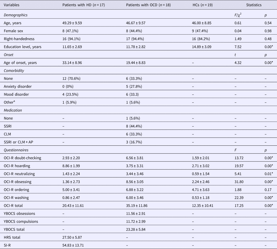
Sociodemographic and clinical characteristics of the three groups are presented in Table 1. Besides the anticipated across-group differences in clinical scores, patient groups differed in age at onset, and also differed from HCs in years of education.
Sample demographic and clinical information

Mean and standard deviation are provided for continuous variables, whereas number of cases and percentage are presented for categorical variables. F- and t tests were performed for continuous variables, while χ2 tests were used for categorical variables.
*Denotes significant between-group differences (p < 0.05).
a One patient of the HD group had a previous history of alcohol abuse and one patient of the OCD group had a previous history of pathological gambling.
CLM, Clomipramine; HRS, Hoarding Rating Scale; OCD, obsessive-compulsive disorder; OCI-R, Obsessive-Compulsive Inventory-Revised; SI-R, Saving Inventory-Revised; SSRI, selective serotonin reuptake inhibitors; SSRI or CLM + AP, selective serotonin reuptake inhibitors or Clomipramine potentiated with an antipsychotic; YBOCS, Yale-Brown Obsessive-Compulsive Scale.
Behavioral results
In the stop-signal task, the HD group showed a lower mean RT in correct go-trials and committed more errors in no-go trials (i.e. commission errors) when compared to OCD and HCs. During the switch-signal task, OCD patients showed fewer correct go trials than HCs, while the HD group showed a lower mean RT in correct go-trials in comparison with OCD patients and fewer correct go trials than HCs. Likewise, they committed more errors than the other two groups (more commission errors than the OCD group and more commission and omission errors than HCs). These results are presented in online Supplementary Table S1 and Fig. S2.
fMRI results
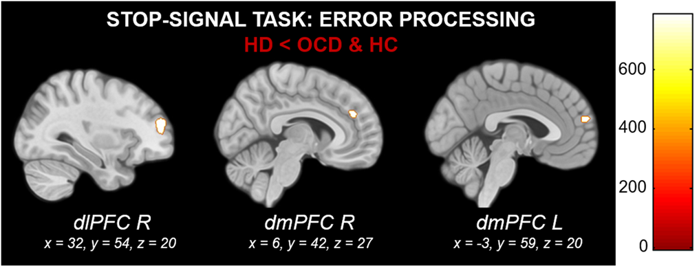
In the stop-signal task, patients with HD exhibited decreased activation of the right dlPFC and the bilateral dmPFC during error processing in comparison with patients with OCD and HCs. No across-group differences were observed in the inhibition contrast. These results are presented in Fig. 1 and online Supplementary Table S2.
Between-group differences in brain activation during error processing in the stop-signal task. Color bar indicates TFCE values, which result from combining voxel-height and cluster size. HD: hoarding disorder; OCD: obsessive-compulsive disorder; HC: healthy controls; dlPFC: dorsolateral prefrontal cortex; dmPFC: dorsomedial prefrontal cortex; R: right; L: left.

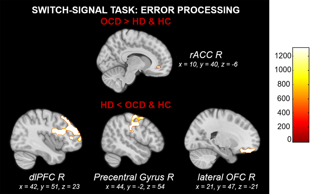
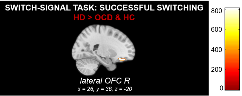
In the switch-signal task, OCD patients showed increased activation of the right rostral ACC (rACC) during error processing compared to HD and HCs. By contrast, HD patients exhibited a right-lateralized decreased activation of the dlPFC, the precentral gyrus and the lateral OFC in comparison with both OCD and HCs during error processing (see Fig. 2). Finally, HD patients showed greater activation in the right lateral OFC during switching compared to OCD and HCs (see Fig. 3). These results are summarized in online Supplementary Table S2.
Between-group differences in brain activation during error processing in the switch-signal task. Color bar indicates TFCE values, which result from combining voxel-height and cluster size. HD: hoarding disorder; OCD: obsessive-compulsive disorder; HC: healthy controls; dlPFC: dorsolateral prefrontal cortex; OFC: orbitofrontal cortex; rACC: rostral anterior cingulate cortex. R: right.

Between-group differences in brain activation during successful switching in the switch-signal task. Color bar indicates TFCE values, which result from combining voxel-height and cluster size. HD: hoarding disorder; OCD: obsessive-compulsive disorder; HC: healthy controls; OFC: orbitofrontal cortex; R: right.

Whole-brain level analyses are presented in online Supplementary Figs S3 and S4 and Table S3. To summarize, these analyses showed significant hypoactivations in the HD group during error processing involving the right somatosensory cortex during stop-signal, and posterior temporo-occipital and cerebellar regions during the switch-signal task.
All the above findings remained significant when controlling for years of education. Moreover, further statistical analyses were performed to assess for potential effects of comorbidity on our findings, finding no significant differences in brain activation results between patients with and without comorbidities. Medication effects were evaluated within the OCD group and we also found no differences when comparing patients with different treatments.
Correlations of brain activity with behavioral and clinical measures
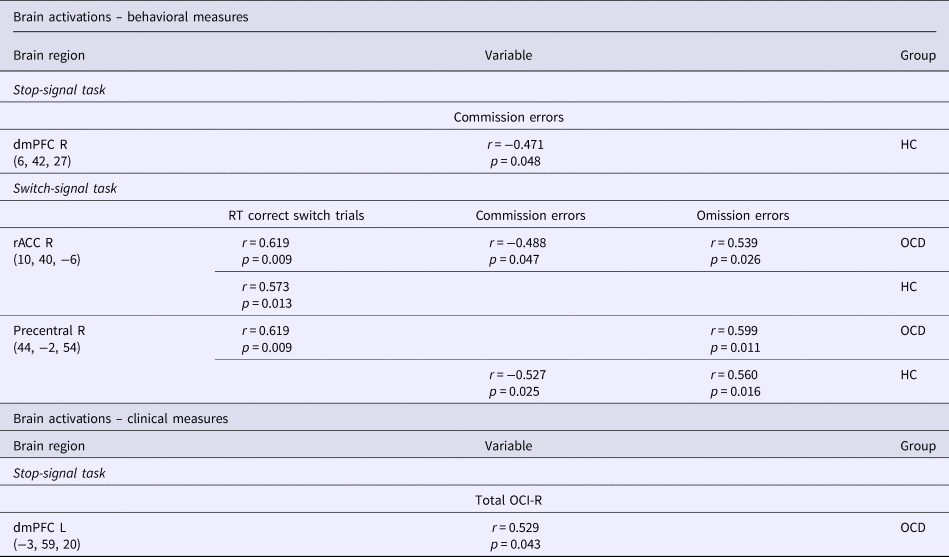
Regarding brain activation estimates extracted from the stop-signal task, a significant negative correlation was observed between right dmPFC activation during error processing and the number of commission errors in the HC group.
As for the switch-signal task (error processing), right precentral gyrus activation was positively associated with RT in correct switch trials and the number of omission errors in the OCD group. Similarly, right precentral gyrus activation was also positively associated with the number of omission errors and negatively associated with the number of commission errors in the HC group. Finally, right rACC activation was positively correlated with RT in correct switch trials in the OCD and HD groups. Additionally, it was negatively associated with the number of commission errors and positively correlated with the number of omission errors in the OCD group.
Concerning clinical measures, left dmPFC activation during the stop-signal task correlated positively with the total OCI-R score in the OCD group.
These results are presented in Table 2 and online Supplementary Fig. S5. Correlations between outside frontal lobe findings and behavioral and clinical measures are displayed in online Supplementary Table S4.
Significant correlations of brain activity with behavioral and clinical measures

Correlation coefficients (r) and p-values (p) are provided for each correlation.
dmPFC, dorsomedial prefrontal cortex; rACC, rostral anterior cingulate cortex; L, left; R, right; OCD, obsessive-compulsive disorder; HC, healthy controls; RT, mean reaction time; OCI-R, Obsessive-Compulsive Inventory-Revised.
Discussion
In the current study, we examined behavioral and brain activation differences in response inhibition, switching and error processing across three different groups of participants: OCD, HD and HCs. The results of this research allow for characterizing the differential neurocognitive profile of the patient groups in a series of executive domains ostensibly important for understanding clinical features, as well as for substantiating the division between HD and OCD in neurobiological terms. Subjects with OCD showed fewer successful go trials than HCs during response switching, which was accompanied by an increased activation of the right rACC during error processing. Conversely, subjects with HD showed shorter RTs in correct go-trials and made more errors during both the stop- and the switch-signal tasks. This was accompanied by an increased activation of the right lateral OFC during response switching and a frontal hypoactivation during error monitoring compared to OCD and HC groups.
Response inhibition and switching depend on prefrontal (including dlPFC, vlPFC and ACC), parietal and striatal regions (Hedden and Gabrieli, Reference Hedden and Gabrieli2010; Zhang et al., Reference Zhang, Geng and Lee2017). Previous studies have shown that these processes are impaired in OCD (Snyder et al., Reference Snyder, Kaiser, Warren, Heller, Hall and Hospital2015), and recent meta-analyses assessing inhibitory control in OCD reported functional alterations partially overlapping with these areas (Norman et al., Reference Norman, Carlisi, Lukito, Hart, Mataix-Cols, Radua and Rubia2016, Reference Norman, Taylor, Liu, Radua, Chye, De Wit, Huyser, Karahanoglu, Luks, Manoach, Mathews, Rubia, Suo, van den Heuvel, Yücel and Fitzgerald2018). In the current study, however, no significant differences in stop-signal performance or associated brain activations were observed between OCD and HCs. Despite previous reports of response inhibition deficits (Snyder et al., Reference Snyder, Kaiser, Warren, Heller, Hall and Hospital2015; Norman et al., Reference Norman, Carlisi, Lukito, Hart, Mataix-Cols, Radua and Rubia2016), our findings agree with other research where such impairments were not observed (Blom et al., Reference Blom, Samuels, Grados, Chen, Bienvenu, Riddle, Liang, Brandt and Nestadt2011; Tolin et al., Reference Tolin, Witt and Stevens2014; Hough et al., Reference Hough, Luks, Lai, Vigil, Guillory, Nongpiur, Fekri, Kupferman, Mathalon and Mathews2016). Likewise, in another meta-analysis it was reported that inhibition impairments are not always observed in OCD samples (Shin et al., Reference Shin, Lee, Kim and Kwon2014).
By contrast, during response switching, patients with OCD completed fewer successful go trials than HCs. The analysis of concurrent neurobiological data provides relevant input to the interpretation this finding. In response switching, patients with OCD showed an increased activation of the right rACC during error processing. Moreover, such rACC response correlated positively with RT in correct switch trials and omission errors, and negatively with commission errors. In the context of the well-established role of the ACC in conflict monitoring (Taylor et al., Reference Taylor, Stern and Gehring2007; Hoffmann and Beste, Reference Hoffmann and Beste2015), our results seem to indicate that activation of the right rACC contributes to prevent unpremeditated responses in conflicting scenarios, and agree with previous studies suggesting that as tasks become more demanding, individuals with OCD tend to hyperactivate prefrontal and ACC areas to increase control over responses (Ursu et al., Reference Ursu, Stenger, Shear, Jones, Carter, Katherine Shear, Jones, Carter, Shear, Jones and Carter2003; Maltby et al., Reference Maltby, Tolin, Worhunsky, O'Keefe and Kiehl2005; Yücel et al., Reference Yücel, Harrison, Wood, Fornito, Wellard, Pujol, Clarke, Phillips, Kyrios, Velakoulis and Pantelis2007). Hence, the impaired performance observed during task-switching (i.e. less successful go trials) may likely relate to an excessive error monitoring during the more demanding task.
Regarding patients with HD, they performed significantly worse than HCs and patients with OCD in both tasks. Their pattern of response differed from that observed in OCD, and included shorter RTs and more commission errors, concurring with previous descriptions linking HD with attentional and motor impulsivity, urgency and lack of perseverance (Timpano et al., Reference Timpano, Rasmussen, Exner, Rief, Schmidt and Wilhelm2013). At a neurobiological level, individuals with HD also differed from OCD by showing a widespread pattern of prefrontal hypoactivation when committing errors during both tasks, which is in clear contrast with the rACC hyperactivation observed in OCD. Such a pattern of deactivation is likely to support the under-responsiveness to salient stimuli, as well as the poor insight and cognitive control described in individuals with HD (Tolin et al., Reference Tolin, Witt and Stevens2014; Grisham and Baldwin, Reference Grisham and Baldwin2015). Moreover, in HCs, but not in HD, activation of the right dmPFC was negatively associated with the number of commission errors, indicating that it might contribute to the successful processing of errors and the consequent performance adjustment in an attempt to commit fewer mistakes in subsequent trials. In the same vein, a recent event-related potential (ERP) study found that HD patients exhibited reduced error-related negativity (ERN) (Mathews et al., Reference Mathews, Perez, Roach, Fekri, Vigil, Kupferman and Mathalon2016). ERN is a frontocentrally distributed ERP component that peaks after conscious appraisal of errors committed during RT tasks. However, in the case of unapprised errors, which may be the case of individuals with HD, ERN appears reduced.
In addition, the prefrontal hypoactivation observed in HD extended to the precentral gyrus. Functional alterations surrounding the primary motor region are not unprecedented; however, increases rather than decreases have been reported in HD compared to OCD and HCs (Saxena et al., Reference Saxena, Brody, Maidment, Smith, Zohrabi, Katz, Baker and Baxter2004; Tolin et al., Reference Tolin, Witt and Stevens2014; Grisham and Baldwin, Reference Grisham and Baldwin2015; Hough et al., Reference Hough, Luks, Lai, Vigil, Guillory, Nongpiur, Fekri, Kupferman, Mathalon and Mathews2016). Importantly, however, in our study, right precentral gyrus activation seems to lead to longer RT and more omission errors, and also to fewer commission errors, in HC and OCD groups, but not in HD. Such findings may indicate that activation of the motor cortex plays a role in response inhibition, and, consequently, hypoactivation of the primary motor region might also contribute to the impulsive pattern of behavior described above. Further, such hypoactivation extended to other brain areas outside the frontal lobe, including primary somatosensory and visual processing regions, reflecting a widespread decrease in brain activity during error feedback. This is likely to lead to diminished attention to error signals and a reduced use of this information to steer subsequent behavior.
Patients with HD also displayed an increased activation of the right lateral OFC during successful response switching. Previous studies have reported increased activations of the OFC in individuals with HD when taking discarding decisions (An et al., Reference An, Mataix-Cols, Lawrence, Wooderson, Giampietro, Speckens, Brammer and Phillips2009; Tolin et al., Reference Tolin, Kiehl, Worhunsky, Book and Maltby2009). A similar finding was observed during a go/no-go paradigm in comparison with HCs (Hough et al., Reference Hough, Luks, Lai, Vigil, Guillory, Nongpiur, Fekri, Kupferman, Mathalon and Mathews2016), although opposite findings have also been reported using the same protocol in comparison with patients with OCD (Tolin et al., Reference Tolin, Witt and Stevens2014). Such mixed findings might be explained by the fact that these different protocols rely on multiple processes such as response inhibition, stimulus-reinforce associations, or the representation of reward and punishment value, which have been broadly associated with OFC function. However, such processes may be mapped onto different regions across the medial-lateral and rostral-caudal axes of the OFC (Kringelbach and Rolls, Reference Kringelbach and Rolls2004). In this sense, recent proposals suggest that the core function of lateral OFC might be the formation and modification of cognitive maps defining task space (Stalnaker et al., Reference Stalnaker, Cooch and Schoenbaum2015). This may imply that the lateral OFC might not be strictly necessary for any of the above functions unless the formation of a cognitive map is required, most likely during the performance of cognitive demanding tasks. Therefore, patients with HD might overly activate the right lateral OFC in an attempt to recruit cognitive resources for successful response switching.
This study is not without limitations. Although we had enough statistical power to detect significant findings after correction for multiple testing, given our limited sample size our results should be described as preliminary, and further research is warranted to better characterize the differences and overlaps between OCD and HD in these and other protocols. Apart from sample size, our clinical groups differed in terms of age at onset, although such difference responds to the specific clinical features of the disorders and confer external validity to our study. Moreover, clinical groups differed from HCs at the education level. However, explicit testing reported no significant effects of this variable. Likewise, the presence of comorbidities in patient groups did not affect our findings. The use of psychotropic medication seemed to also have null effects on our results, although it was not possible to control for this variable across groups because it only applied to patients with OCD. Although given at stable doses throughout the study, medication effects should be controlled for in future studies. Finally, as in all correlational studies, we cannot infer causality from our results. In consequence, caution is warranted when interpreting the associations between brain activation and behavioral or clinical features.
In summary, our results indicate that patients with OCD only differ from HCs in the hyperactivation of the rACC observed during error processing in task switching, suggesting an increased error monitoring in demanding settings. Conversely, patients with HD differ from OCD in showing a widespread deactivation during error processing in response inhibition and task switching. Likewise, they also differ from OCD in displaying an abnormal hyperactivation of the right lateral OFC during response switching, which can be related to the need of recruiting more cognitive resources for successful switching in the context of limited error processing and an impulsive pattern of response. Our study shows that OCD and HD differ in their neurocognitive and neurobiological profiles during executive performance, substantiating the clinical separation between these disorders. The results reported here may eventually be used as objective biomarkers in studies and clinical trials aiming at discriminating between these disorders.
Supplementary material
The supplementary material for this article can be found at https://doi.org/10.1017/S0033291719000515
Financial support
This study was supported in part by the Carlos III Health Institute (PI13/01958, PI14/00413, PI16/00950, PI16/00889 and CIBER-CB06/03/0034); FEDER funds (‘A way to build Europe’) and by the Agency of University and Research Funding Management of the Catalan Government (2014SGR1672). M.S. is supported by a CIBERSAM Ph.D. grant (CNV665/914), IM-Z is supported by a P-FIS grant from the Carlos III Health Institute (FI17/00294), MP-P is supported by a FI Grant from AGAUR-Generalitat de Catalunya (2015 FI_B 00839) and C.S.-M. is supported by a Miguel Servet contract (CPII16/00048) from the Carlos III Health Institute.
Conflict of interest
None.