Introduction
Since the 1900s, numerous trypanosome species have been identified worldwide, infecting all classes of vertebrates, with the majority appearing to cause no disease to their hosts (Hoare, Reference Hoare1972; Tyler and Engman, Reference Tyler and Engman2001; Hamilton et al., Reference Hamilton, Gibson and Stevens2007). Bats (order: Chiroptera) evolved ~50–52 million years ago during the early Eocene period and comprise ~20% of extant mammals (Teeling et al., Reference Teeling, Springer, Madsen, Bates, O'brien and Murphy2005). Bats are among the most common hosts of a large variety of trypanosome species worldwide, with the subgenus Schizotrypanum comprising the majority of the trypanosomes reported in bats throughout South America, Europe, Africa and Asia (Hamilton et al., Reference Hamilton, Teixeira and Stevens2012b; Lima et al., Reference Lima, Espinosa-Alvarez, Hamilton, Neves, Takata, Campaner, Attias, de Souza, Camargo and Teixeira2013; Mafie et al., Reference Mafie, Rupa, Takano, Suzuki, Maeda and Sato2018; Wang et al., Reference Wang, Han, Zhao, Liu, Luo, Wen, Qin, Zhou, Qi, Yu and Yu2019). Trypanosomes in Chiroptera were first described in Miniopterus schreibersii from Italy by Dionisi in 1899 (Baker et al., Reference Baker, Green, Chaloner and Gaborak1972). Since then over 30 species of trypanosomes have been reported in more than 100 bat species, including well defined Trypanosoma cruzi, Trypanosoma vespertilionis, Trypanosoma rangeli and the globally distributed Trypanosoma dionisii (Beltz, Reference Beltz2017).
Relatively little is known about Trypanosoma species in native Australian bat species. Trypanosoma pteropi was the first described trypanosome in Pteropus sp. from north Queensland (Breinl, Reference Breinl1913). Similar trypanosomes were characterized by Mackerras (Reference Mackerras1959) in Pteropus gouldii, however, they were described as smaller in length, had a pointed anterior and a rounded kinetoplast, similar to T. vespertilionis from European bats, which shares biological characteristics with T. cruzi with both species having the ability to reproduce in organs (Mackerras, Reference Mackerras1959). Mackerras (Reference Mackerras1959) also described a very tiny and slender trypanosome named Trypanosoma hipposideri from Hipposideros ater captured in northern Queensland. It was reported to be smaller than T. vespertilionis and T. pteropi, with a large distinctive kinetoplast and delicate short free flagellum. More recently, the first molecular study identified a high prevalence of Trypanosoma vegrandis in two species of pteropid bats (Pteropus alecto and Pteropus scapulatus) and two species of microbats (Chalinolobus gouldii and Nyctophilus geoffroyi) from Western Australian (Austen et al., Reference Austen, O'Dea, Jackson and Ryan2015). Trypanosoma teixeirae from a native Australian bat, P. scapulatus was also recently described by Mackie et al. (Reference Mackie, Stenner, Gillett, Barbosa, Ryan and Irwin2017) and further characterized by Barbosa et al. (Reference Barbosa, Mackie, Stenner, Gillett, Irwin and Ryan2016). This trypanosome is longer in length than both T. pteropi and T. hipposideri and genetically similar to Trypanosoma rangeli and Trypanosoma minasense (Mackie et al., Reference Mackie, Stenner, Gillett, Barbosa, Ryan and Irwin2017). Interestingly T. teixeirae is the first bat trypanosome in Australia to cause clinical disease in its host, with descriptions of icterus and anaemia (Mackie et al., Reference Mackie, Stenner, Gillett, Barbosa, Ryan and Irwin2017).
The aim of the present study was to investigate trypanosome species in seven species of microbats from the South-West Botanical Province of Western Australia, to gain insight into the prevalence and genetic diversity of trypanosomes in these species.
Methods
Field sample collection
Animal work was performed with approval from the Murdoch University Animal Ethics Committee under permit #R2882/16. Fieldwork was conducted between the 30th of November to the 9th of December 2016 at Charles Darwin Reserve (−29.581817, 117.001820) and Mount Gibson Wildlife Sanctuary (−29.630577, 117.277211), located within the south-west region of Western Australia, a globally recognized biodiversity hotspot (Myers et al., Reference Myers, Mittermeier, Mittermeier, da Fonseca and Kent2000). Bats were caught using harp traps (Faunatech, Australia) and mist nets (Ecotone, Australia) set over water points at sunset. The mist nets were monitored constantly with bats removed from the nets immediately after capture. Harp traps were left unmonitored for approximately 3 hours and then checked for individuals captured in the protective bagging.
Sampling occurred at the capture site with no individuals being held for greater than 2 hours and handling limited to 5 min per individual to minimize stress. Palpably pregnant females were released without sampling. Bats were identified to species level using published keys (Churchill, Reference Churchill2008). Blood samples were collected from 86 microbats representing 7 known species; Chalinolobus gouldii (n = 53), Chalinolobus morio (n = 3), Nyctophilus geoffroyi (n = 16), Nyctophilus major (n = 1), Ozimops sp. (n = 2), Scotorepens balstoni (n = 7) and Vespadelus baverstocki (n = 4). A measure of 10 μL of blood was collected from the uropatagial vein using a 25-gauge needle and a micropipette, and then diluted in sterile 1:10 phosphate buffered saline. Individual blood smears were made, air dried and fixed in 100% methanol. Blood samples were stored at 4°C in the field and later stored at −20°C until use.
DNA extraction and molecular analysis
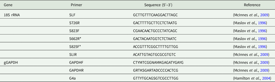
DNA was extracted from ethylene diamine tetraacetic acid (EDTA)-preserved whole blood using a NucleoMag VET kit (Macherey-Nagel, Germany) on an automated MagMax 96 processor (Life Technologies Corporation, USA) according to the manufacturer's instructions. Partial fragments of Trypanosoma spp. DNA were amplified from the 18S rDNA and gGAPDH loci using primers previously described by Maslov et al. (Reference Maslov, Lukes, Jirku and Simpson1996) and McInnes et al. (Reference Mcinnes, Gillett, Ryan, Austen, Campbell, Hanger and Reid2009), (Table 1). Initially, samples were screened using a single round polymerase chain reaction (PCR) to amplify a small fragment (~350 bp) of Trypanosoma DNA using the 18S rDNA primer set S825F and S662R. Once sequences were obtained, a subsample of positives representing different genotypes in the initial targeted PCR assay, were re-amplified to obtain a larger product size using the nested and hemi nested 18S rDNA and gGAPDH primer sets, respectively. In addition, bats were also screened for T. vegrandis as previously described by Austen et al. (Reference Austen, O'Dea, Jackson and Ryan2015).
Primers used for Trypanosoma PCR

*Denotes primer combination used for initial screening of the short 18S rRNA fragment.
The 18S rDNA PCR assays were performed in 25 μL volumes and contained approximately 50 ng of gDNA, 0.4 μ m of each primer and 12.5 μL GoTaq® Green master mix (Promega, USA). A 1 μL aliquot of the primary PCR product was used as the DNA template for the nested round of PCR. All PCR cycling conditions consisted of an initial cycle of denaturation at 95°C for 5 min, 50°C for 2 min, 72°C for 4 min followed by 40 cycles of denaturation at 94°C for 30 sec, annealing at 52°C (primary reaction) or 55°C (secondary reaction) for 30 sec and extension at 72°C for 2.20 min (primary reaction) or 1 min (secondary), followed by a final extension at 72°C for 7 min. The PCR product was run on a 1.5% agarose gel stained with SYBR safe (Invitrogen, USA) and viewed under UV illumination. Amplified PCR products of the appropriate size were excised from the gel using a sterile scalpel blade for each band to prevent cross-contamination and purified using an in-house filter tip method and used for sequencing without any further purification as previously described (Yang et al., Reference Yang, Murphy, Song, Ng-Hublin, Estcourt, Hijjawi, Chalmers, Hadfield, Bath, Gordon and Ryan2013). All PCR amplicons were submitted to the Australian Genome Research Facility (Perth, Western Australia) for unidirectional sequencing. Sequences were imported and trimmed in Geneious v10 (Kearse et al., Reference Kearse, Moir, Wilson, Stones-Havas, Cheung, Sturrock, Buxton, Cooper, Markowitz, Duran, Thierer, Ashton, Meintjes and Drummond2012) and then subject to BLAST analysis using BLASTN 2.10.0 + (Zhang et al., Reference Zhang, Schwartz, Wagner and Miller2000) against nucleotide collection (nt) database (Morgulis et al., Reference Morgulis, Coulouris, Raytselis, Madden, Agarwala and Schäffer2008) to identify most similar species and genotypes.
Prevalence
The prevalence of trypanosomes in bat blood samples from each host species was calculated based on the number positive at the 18S rDNA locus, with 95% confidence intervals calculated assuming a binomial distribution using the software Quantitative Parasitology 3.0 (Rozsa et al., Reference Rozsa, Reiczigel and Majoros2000).
Phylogenetic analysis
Reference sequences for members within the T. cruzi clade were retrieved from GenBank (Benson et al., Reference Benson, Cavanaugh, Clark, Karsch-Mizrachi, Lipman, Ostell and Sayers2017) (available in Supplementary Table 1) and aligned with sequences obtained in the present study using MUSCLE (Edgar, Reference Edgar2004). The 18S rDNA alignment was then curated using gblocks (Castresana, Reference Castresana2000) with less stringent parameters (only reference sequences >1 kb were retained for phylogenetic analysis). A 1,350 bp region of the 18S rDNA locus and a 674 bp region of the gGAPDH locus were then used for phylogenetic purposes. Phylogenies were inferred using the maximum likelihood (ML) and Bayesian inferences (BI). The ML analysis was conducted in MEGA 7 (Kumar et al., Reference Kumar, Stecher and Tamura2016), the optimal model was chosen using the model selection feature. ML phylogenies were conducted using the General Time Reversible (GTR) (Nei and Kumar, Reference Nei and Kumar2000) + Gamma (G) (0.05) model for 18S rDNA and concatenated 18S rDNA and GAPDH sequences (2024 bp) while the Tamura 3-parameter (TN92) (Tamura, Reference Tamura1992) + G (0.29) model was used for gGAPDH, with 5,000 bootstrap replicates. The BI analysis was conducted in MrBayes v3.2.6 (Ronquist et al., Reference Ronquist, Teslenko, van der Mark, Ayres, Darling, Hohna, Larget, Liu, Suchard and Huelsenbeck2012), using the GTR + G model for both genes with a Markov chain Monte Carlo length of 1 10 000, subsampling every 200 iterations and a burn-in rate of 25%. The reptile trypanosome, Trypanosoma varani was used as an outgroup in all analyses.
Morphological analysis
Thin blood smears were stained with Modified Wright–Giemsa (Hematek® Stain Pak) using a Hema-Tek SlideStainer (Ames Company Division, Miles Laboratories Pty Ltd., Springvale Victoria, Australia) and examined for the presence of trypanosomes. The slides were mounted using DPX neutral mounting medium (LabChem, Victoria, Australia) and a coverslip applied. Digital light micrograph images of trypomastigotes were taken at × 1000 magnification using Image-Pro Express software (Media Cybernetics, Inc., Bethesda, Maryland, USA). Morphological measurements including total length (TL: length of body measured along the mid-line including free flagellum), breadth (B: maximum breadth measured at the level of the nucleus including undulating membrane), kinetoplast to nucleus (KN: distance between the kinetoplast and posterior edge of the nucleus), nucleus to anterior (NA: distance between the anterior edge of the nucleus and the anterior end of the body), posterior to the kinetoplast (PK: distance between the posterior end and the kinetoplast) and free flagellum (FF: length of the free flagellum) were measured using Image J (Schneider et al., Reference Schneider, Rasband and Eliceiri2012) and the mean, standard error (±) and range calculated.
Results
Prevalence of trypanosomes in microbats
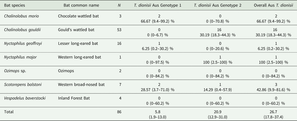
PCR (gel image provided in Supplementary material, Fig. S1), sequencing and phylogenetic analysis of partial consensus DNA fragments of the 18S rDNA and gGAPDH locus identified T. dionisii in Australian microbats. The overall prevalence of T. dionisii in microbats was 26.7% (23/86) with T. dionisii detected in C. gouldii, C. morio, N. geoffroyi, N. major and S. balstoni. No trypanosomes were detected in Ozimops. sp or V. baverstocki. Sequencing and phylogenetic analysis identified two different genotypes named T. dionisii genotype Aus 1 and T. dionisii genotype Aus 2 and were differentiated by the presence of two single nucleotide polymorphisms (SNPs) over the V7 hypervariable region (~350 bp) of the 18S rDNA loci. Genotype Aus 2 was the most prevalent with 20.9% (18/86) of the bats infected, while genotype Aus 1 was less prevalent and infected 5.8% (5/86) of bats. Chalinolobus morio and N. geoffroyi were only infected with genotype Aus 1, while C. gouldii and N. major were only infected with genotype Aus 2. Both Aus genotypes 1 and 2 were detected in S. balstoni (Table 2). Trypanosoma vegrandis was not detected in any of the bats screened by PCR and microscopy.
Prevalence of T. dionisii in microbats from Charles Darwin Reserve and Mount Gibson Reserve, Western Australia

95% confident intervals in brackets.
Phylogenetic analysis
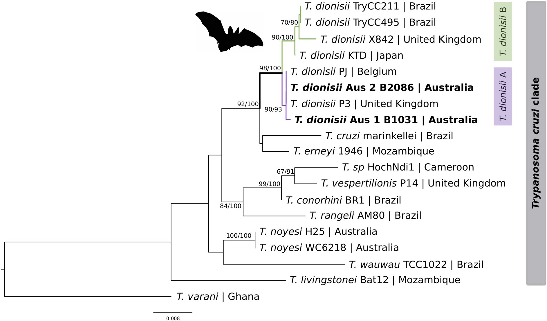
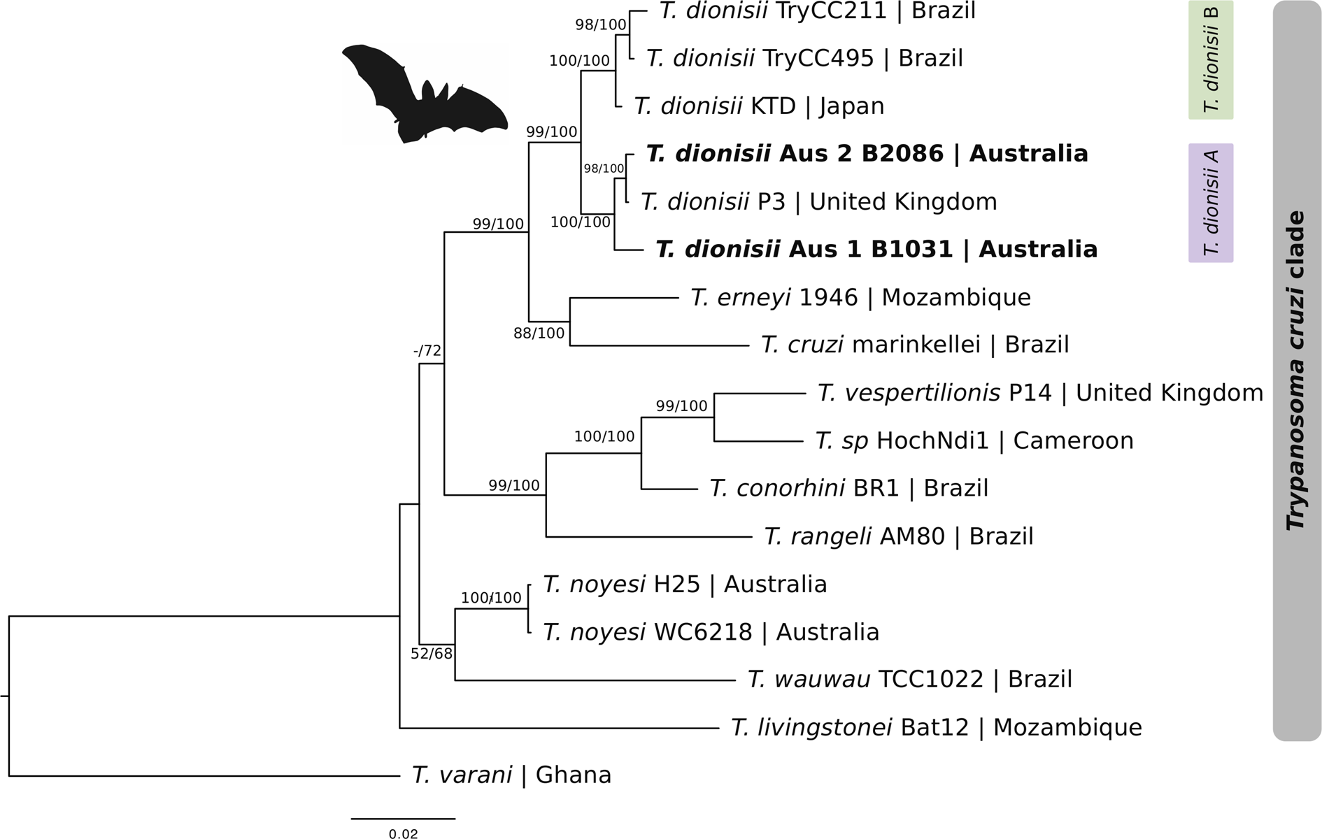
The phylogenetic relationship of the Australian T. dionisii to other Trypanosoma species at the 18S rDNA (Fig. 1), gGAPDH (Fig. 2) and concatenated 18S rDNA and gGAPDH (Fig. 3) sequenced regions, showed similar tree topologies. The two Australian T. dionisii genotypes at the 18S rDNA locus were 99.9% similar to one another and exhibited 3 single nucleotide polymorphisms (SNP's) across 1459 bp of sequence. The Australian T. dionisii genotypes clustered within the T. cruzi clade and grouped together with other T. dionisii isolates including TryCC 211 (FJ001666) from Brazil, KTD (LC326397) from Japan, P3 (AJ009151) from UK (UK) and PJ (AJ009152) from Belgium. Both Australian genotypes 1 and 2 showed a close association with the UK (P3) and Belgium (PJ) isolates, clustering within the T. dionisii clade A. Trypanosoma dionisii genotype Aus 1 exhibited 99.9% similarity to trypanosome isolates PJ and P3 and T. dionisii genotype Aus 2 exhibited a 100% similarity to PJ and P3 isolates from Pipistrellus pipistrellus European bats. The partial 18S rDNA sequences generated in the present study have been submitted to GenBank under accession MT533284 (T. dionisii genotype Aus 1) and MT533287 (T. dionisii genotypes Aus 2).
Maximum likelihood (ML) phylogenetic reconstruction of the Trypanosoma cruzi clade based on a 1350 bp alignment of the 18S rDNA locus. Node numbers correspond to ML and Bayesian inference (BI) support values, respectively (ML values <60 are hidden). Number of substitutions per nucleotide position is represented by the scale bar. Sequences generated in the present study are in bold. GenBank accession numbers for sequences are available in Supplementary Table 1.

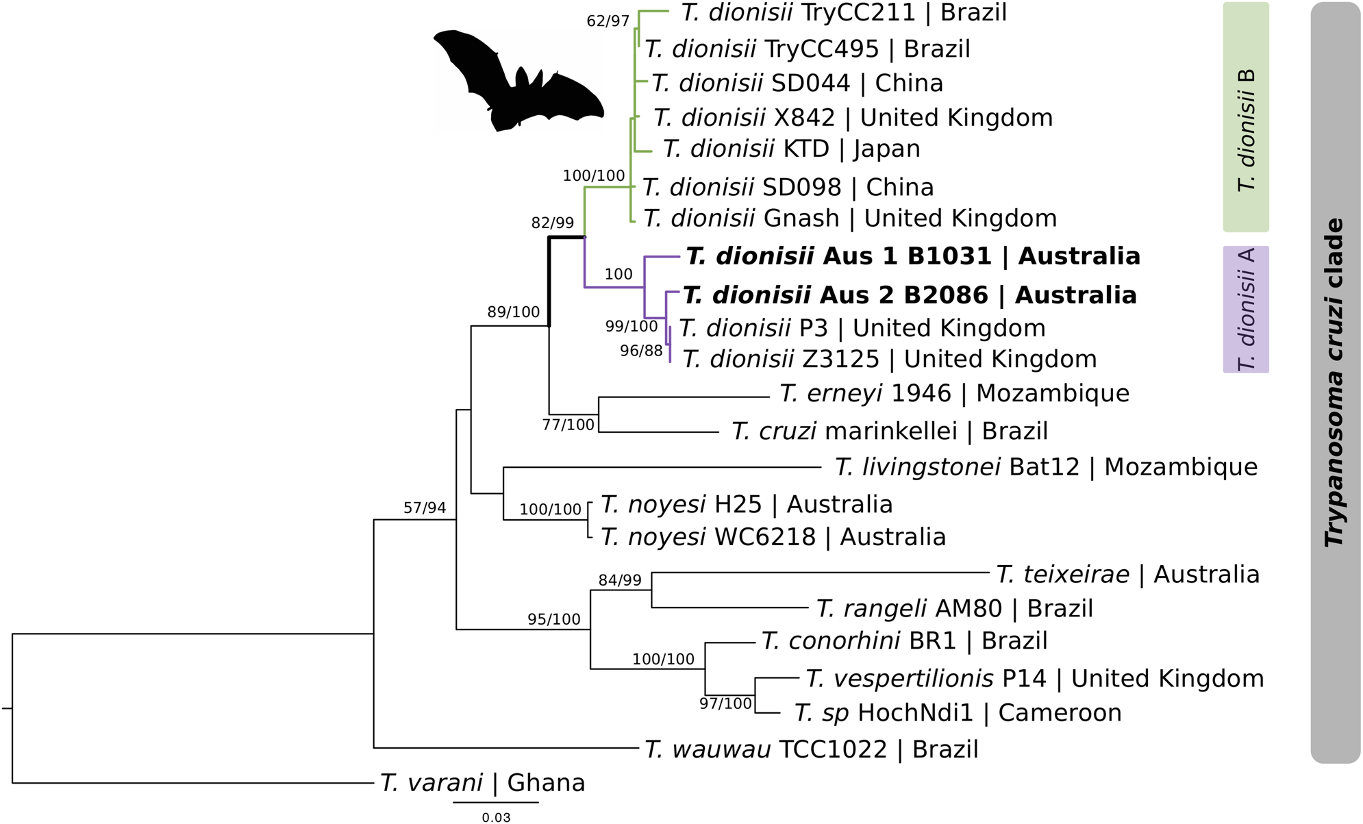
Maximum likelihood (ML) phylogenetic reconstruction of the Trypanosoma cruzi clade based on a 674 bp alignment of the glycosomal glyceraldehyde phosphate dehydrogenase (gGAPDH) gene. Node numbers correspond to ML and Bayesian inference (BI) support values, respectively (ML values <60 are hidden). Number of substitutions per nucleotide position is represented by the scale bar. Sequences generated in the present study are in bold. GenBank accession numbers for sequences are available in Supplementary Table 1.

Maximum likelihood (ML) phylogenetic reconstruction of the Trypanosoma cruzi clade based on a concatenated 18S rRNA and GAPDH sequences. Node numbers correspond to ML and Bayesian inference (BI) support values, respectively (ML values <60 are hidden). Number of substitutions per nucleotide position is represented by the scale bar. Sequences generated in the present study are in bold. GenBank accession numbers for sequences are available in Supplementary Table 1.

At the gGAPDH locus, the Australian T. dionisii genotypes exhibited a 97.6% homology to one another with 16 SNPs across 674 bp of gGAPDH sequence. The Trypanosoma tree topology for gGAPDH, was similar to the 18S rDNA phylogeny, with the Australian T. dionisii genotypes grouping together within the T. dionisii clade with isolates from China (MH393936), UK (FN599056), Brazil (GQ140362), and Japan (LC326399). The T. dionisii genotypes Aus 1 and Aus 2 were most homologous to T. dionisii isolate Z3126 (FN599054) from an English bat (Pipistrellus pygmaeus) with genetic similarities of 97.9% and 99.4%, respectively. The partial gGAPDH sequences generated in the present study have been submitted to GenBank under accession MT536207 (T. dionisii genotype Aus 1) and MT536209 (T. dionisii genotypes Aus 2). The Trypanosoma tree topology for the concatenated sequences produced similar results to both the 18S rDNA and gGAPDH phylogenies, with the Australian T. dionisii genotypes grouping together within the T. dionisii A clade, together with T. dionisii P3 isolate from the UK. Unfortunately, no 18S rDNA and gGAPDH sequences were available for the T. dionisii European isolates Z3126 and PJ, respectively.
Morphology
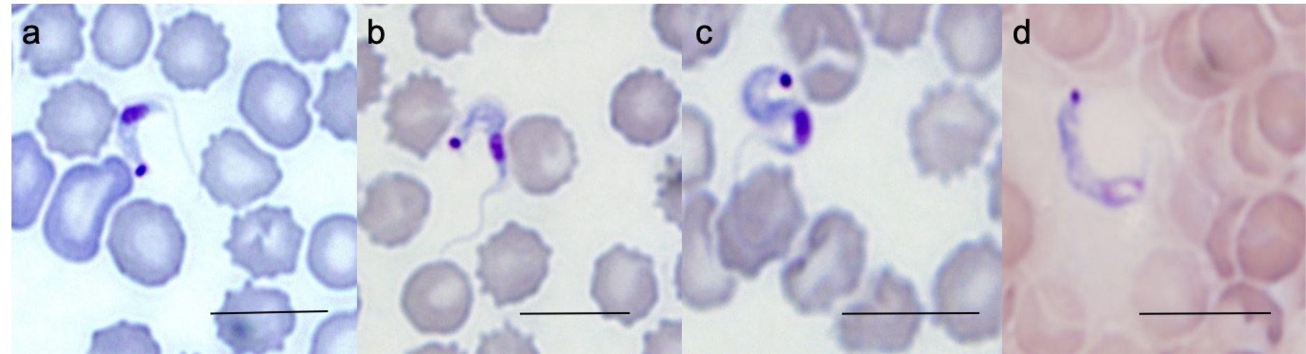
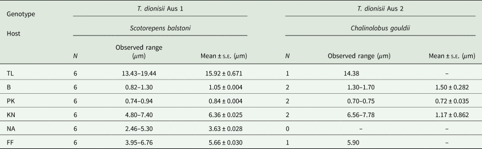
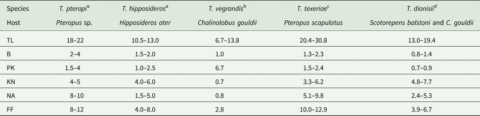
Out of the 23 bats positive for T. dionisii, trypomastigote stages were identified in only 3 bats by blood film analysis. Bat isolate T1-2016-1031 from a S. balstoni microbat (sequence typed as genotype Aus 1) and bat isolate T1-2016-2058 and T1-2016–2102 from C. gouldii microbats (sequence typed as genotype Aus 2) presented with typical trypomastigote stages (Fig. 4). Trypanosoma dionisii showed similar morphology to previously described trypomastigotes from Schizotrypanum bat species. The Australian T. dionisii isolates were slender with a clearly defined rounded kinetoplast, positioned at the posterior end of the trypomastigote, while an elongated nuclear region was located towards the anterior end. The nucleus was positioned at the very anterior tip for T. dionisii genotype Aus 2 isolates and therefore nucleus to anterior distance (NA) measurements could not be determined, as the anterior end was not clearly visible. The undulating membrane was narrow for each of the Aus genotypes and a free flagellum was present at the anterior end of the trypanosome. The morphological parameters were measured for each T. dionisii Aus genotype and are presented in Table 3. Morphometric analysis revealed that trypomastigotes of T. dionisii from Australia are similar to T. hipposideri with overlaps in morphological measurements (Table 4.). There were no significant differences between TL, KA, NA and FF, but T. hipposideri was larger in breadth and PK compared to Australian T. dionisii (P < 0.01).
Trypanosoma dionisii trypomastigotes in Australian microbat blood films. (a–c) Trypomastigotes in blood films from bat isolate T1-2016-1031, Scotorepens balstoni representing genotype Aus 1. (d) Trypomastigote in a blood film from bat isolate T1-2016-2058, Chalinolobus gouldii representing genotype Aus 2. Scale bar represents 10 μ m.

Mean dimensions and standard error (s.e.) of morphological features from Australian Trypanosoma dionisii genotypes Aus 1 and Aus 2, identified in the blood of Australian native microbats

*Morphological abbreviations.
TL, length of body measured along the mid-line including free flagellum (total length); B, maximum breadth measured at the level of the nucleus (including undulating membrane); PK, distance between the posterior end and the kinetoplast; KN, distance between the kinetoplast and posterior edge of the nucleus; NA, distance between the anterior edge of the nucleus and the anterior end of the body; FF, length of the free flagellum.
Number of trypomastigotes measured represented by N, see legend* for terminology.
Morphological comparison of Trypanosoma species isolated from native Australian bats

*Morphological abbreviations.
TL, length of body measured along the mid-line including free flagellum (total length); B, maximum breadth measured at the level of the nucleus (including undulating membrane); PK, distance between the posterior end and the kinetoplast; KN, distance between the kinetoplast and posterior edge of the nucleus; NA, distance between the anterior edge of the nucleus and the anterior end of the body; FF, length of the free flagellum.
Measurements recorded in μ m of trypomastigotes, see legend* for terminology. References:
a Mackerras (Reference Mackerras1959).
b Austen et al. (Reference Austen, O'Dea, Jackson and Ryan2015).
c Barbosa et al. (Reference Barbosa, Mackie, Stenner, Gillett, Irwin and Ryan2016).
d Present study.
Discussion
The present study reports the discovery of T. dionisii in Australia for the first time and is also the first report of this parasite in native Australian bats. Trypanosoma dionisii was identified in five different Australian microbat species, C. gouldii, C. morio, N. geoffroyi, N. major and S. balstoni from the southwest of Western Australia. Vespadelus baverstocki and Ozimops sp were negative for T. dionisii although the small sample sizes of these species are a confounding factor. The high prevalence of 5/7 Australian native bat species infected with T. dionisii indicates low host specificity supporting the cosmopolitan nature of T. dionisii.
Interestingly two different genotypes of T. dionisii were detected in Australia; these genotypes tended to be restricted to individual bat species with C. morio and N. geoffroyi infected with genotype Aus 1 and C. gouldii and N. major infected with genotype Aus 2, whereas S. balstoni was infected with both types. These differences may be due to the opportunistic nature of the sampling since C. gouldii had the highest capture rate of 53 individuals out of the total of 86 individuals (Table 2). To determine the true prevalence of T. dionisii and its genotypes in native Australian bats, larger numbers are needed to reduce sampling bias. Trypanosoma vegrandis was not detected in any of the bats sampled in this study, which is surprising given the high prevalence detected previously by Austen et al. (Reference Austen, O'Dea, Jackson and Ryan2015). These differences may be a result of the geographical location and habitats from which the bats were sourced. The bats from the present study were captured in the Northern wheatbelt region within the Southwest Botanical Province of Western Australia, whereas the T. vegrandis positive bats from Austen et al. (Reference Austen, O'Dea, Jackson and Ryan2015) were sampled from the semi-urban Perth Hills region of Western Australia. These two regions exhibit vastly different climatic conditions and land-use regimes with large areas of habitat fragmentation separating them. Aside from potential environmental or landscape factors, the reason for this discrepancy in prevalence is unknown.
Trypanosoma dionisii is globally distributed and has been described in Chiroptera from North America, South America, Europe, Africa and more recently Asia (Hamilton et al., Reference Hamilton, Teixeira and Stevens2012b; Lima et al., Reference Lima, Espinosa-Alvarez, Hamilton, Neves, Takata, Campaner, Attias, de Souza, Camargo and Teixeira2013; Hodo et al., Reference Hodo, Goodwin, Mayes, Mariscal, Waldrup and Hamer2016; Espinosa-Alvarez et al., Reference Espinosa-Alvarez, Ortiz, Lima, Costa-Martins, Serrano, Herder, Buck, Camargo, Hamilton, Stevens and Teixeira2018; Mafie et al., Reference Mafie, Rupa, Takano, Suzuki, Maeda and Sato2018; Wang et al., Reference Wang, Han, Zhao, Liu, Luo, Wen, Qin, Zhou, Qi, Yu and Yu2019). The discovery of T. dionisii in Australia now broadens its range to include the sixth continent. The prevalence of T. dionisii in Australian bats in the present study was 26.7%; much higher than 9.6% (16/154) reported in bat species from England (Hamilton et al., Reference Hamilton, Cruickshank, Stevens, Teixeira and Mathews2012a) and also in Asia, where a 10.3% (13/126) prevalence was detected in E. serotinus and Myotis pequinius bats from China (Wang et al., Reference Wang, Han, Zhao, Liu, Luo, Wen, Qin, Zhou, Qi, Yu and Yu2019) and 2.1% (2/94) in Miniopterus fuliginosus bats from Japan (Mafie et al., Reference Mafie, Rupa, Takano, Suzuki, Maeda and Sato2018). Only a small percentage of T. dionisii (1.5% −9/593) has been detected in various bat species from Texas, USA (Hodo et al., Reference Hodo, Goodwin, Mayes, Mariscal, Waldrup and Hamer2016).
The geographical origin of the T. cruzi clade is the subject of debate. Currently, the clade is composed of two main sister phylogenetic lineages; one represented by the subgenus Schizotrypanum, harbouring T. cruzi in bats and mammals, and other members of this group (T. cruzi marinkellei, T. erneyi and T. dionisii), all of which are restricted to bats. The second lineage consists of the species T. rangeli, which has a broad mammalian host range, including humans, T. conorhini which infects rodents and T. vespertilionis, restricted to bats (Cavazzana et al., Reference Cavazzana, Marcili, Lima, da Silva, Junqueira, Veludo, Viola, Campaner, Nunes, Paiva, Coura, Camargo and Teixeira2010; Garcia et al., Reference Garcia, Ortiz, Osorio, Torrico, Torrico and Solari2012; Lima et al., Reference Lima, Silva, Neves, Attias, Takata, Campaner, de Souza, Hamilton and Teixeira2012; Espinosa-Alvarez et al., Reference Espinosa-Alvarez, Ortiz, Lima, Costa-Martins, Serrano, Herder, Buck, Camargo, Hamilton, Stevens and Teixeira2018; Wang et al., Reference Wang, Han, Zhao, Liu, Luo, Wen, Qin, Zhou, Qi, Yu and Yu2019). The phylogenetic placement of an Australian marsupial trypanosome T. noyesi (originally known as T. sp. H25) within the T. cruzi clade together with species predominately from South America, prompted a ‘southern supercontinent’ hypothesis by Stevens et al. (Reference Stevens, Noyes and Gibson1998) who proposed that ancestral T. c. cruzi speciated in marsupials after the separation of South America from the Australian continent approximately 40 million years ago (Stevens et al., Reference Stevens, Noyes and Gibson1998). In contrast, Hamilton et al. (Reference Hamilton, Teixeira and Stevens2012b) proposed the ‘bat seeding hypothesis’, which proposes that bats are the ancestral hosts of the T. cruzi clade and that ancestral trypanosomes evolved into terrestrial placental mammalian and marsupial hosts over time, resulting in new linages within the clades (Lima et al., Reference Lima, Silva, Neves, Attias, Takata, Campaner, de Souza, Hamilton and Teixeira2012; Hamilton et al., Reference Hamilton, Teixeira and Stevens2012b). The close genetic relationship of the Australian T. dionisii to T. dionisii isolates PJ, P3 and Z3126 from Europe would seem to support Hamilton's bat seeding theory, with the likelihood that European Old World trypanosomes seeded into Australian mammalian species.
Furthermore, recent studies also show a closer genetic relationship between Australian trypanosomes and those found in other regions of the world, than with each other (Lima et al., Reference Lima, Silva, Neves, Attias, Takata, Campaner, de Souza, Hamilton and Teixeira2012; Hamilton et al., Reference Hamilton, Teixeira and Stevens2012b). For example, Trypanosoma copemani, which infects various Australian marsupials shares genetic similarities with Trypanosoma pestanai from English badgers (Noyes et al., Reference Noyes, Stevens, Teixeira, Phelan and Holz1999; Austen et al., Reference Austen, Jefferies, Friend, Ryan, Adams and Reid2009), while Trypanosoma lewisi isolated from an Australian native rat shares a close genetic relationship to T. lewisi from England (Egan et al., Reference Egan, Taylor, Austen, Banks, Ahlstrom, Ryan, Irwin and Oskam2020). In addition, another trypanosome from an indigenous Australian bird, the currawong (Strepera sp.) was closely related to trypanosomes isolated from European birds and their vectors (Hamilton et al., Reference Hamilton, Stevens, Gidley, Holz and Gibson2005).
A study by Hamilton et al. (Reference Hamilton, Cruickshank, Stevens, Teixeira and Mathews2012a) on T. dionisii from English bats detected two different genotypes which were subdivided into two major clades, with type A found only in English bats and type B identified in bats from Brazil and England. In the present study, the Australian T. dionisii isolates are grouped within the T. dionisii type A described by Hamilton et al. (Reference Hamilton, Cruickshank, Stevens, Teixeira and Mathews2012a), suggesting that T. dionisii from Australian bats has indeed descended from European bat trypanosomes. In addition, the 100% homology of T. dionisii genotype Aus 2 (B2086) and the 99.9% homology of T. dionisii genotype Aus 1 to both PJ and P3 isolates from Belgium and the UK respectively, at the 18S rDNA locus, suggest the movement of this bat trypanosome when bat species began to diverge as the result of the continents breaking away. Unfortunately, no sequence for PJ at the gGAPDH locus was available for phylogenetic comparison, so isolate Z3125 from a UK bat (Pipistrellus pygmaeus) was used instead. As with the 18S rDNA, phylogenetic analysis at the gGAPDH locus showed that genotype Aus 2 exhibited the greatest homology to English bat isolates with 99.4% homology to P3 and Z3126. The T. dionisii genotype Aus 1 was less homologous at 97.9% to the same English isolates. How these ‘English’ bat trypanosomes have established in Australia is uncertain, with the precise route of parasite movement unknown. The grouping of T. dionisii from Asian bats into the T. dionisii B clade (in contrast to the T. dionisii A clade, in which both the Australian T. dionisii genotypes Aus 1 and 2 grouped), complicates the rationale that ancestral bats may have travelled over millennia from Europe via Asia and then into Australia. An alternative scenario is the movement of infected bats across different continents by anthropogenic transport (Hamilton et al., Reference Hamilton, Cruickshank, Stevens, Teixeira and Mathews2012a). For example, a study by Constantine (Reference Constantine2003) cited several reports in which bats have been inadvertently carried across landmasses by humans, by roosting in aeroplanes and/or ships. Presently it is unclear how T. dionisii entered Australia and established in native bat species. Additional sampling, analyses and evolutionary clock modelling would be needed to build up a likely scenario.
From a morphological perspective, Trypanosoma species of the subgenus Schizotrypanum are indistinguishable from each other. They all conform in morphology and in their lifecycles to the type-specimen Trypanosoma (Schizotrypanum) cruzi (Hoare, Reference Hoare1972). They are dimorphic parasites with both slender and broad forms. Typically, their trypomastigotes are relatively small, curved and have a distinctive round to oval kinetoplast. Based on overall morphological characteristics, the trypanosomes of the Australian T. dionisii aligns with the general descriptions of trypomastigotes previously described for the subgenus Schizotrypanum. The Australian isolates have a large distinctive round kinetoplast, are small and slender and range in total length between 13.43–19.44 μ m. In comparison to other described Australian bat trypanosomes, T. dionisii from native Australian bats, are smaller in size than both T. teixeirae and T. pteropi, yet larger and phenotypically different to T. vegrandis. Morphological parameters described for Australian T. dionisii were similar to those of T. hipposideri and fell within or close to the range reported by Mackerras (Reference Mackerras1959). General morphological comparisons between the Australian and Asian T. dionisii isolates could not be determined, as no circulating blood trypomastigotes, [referred to as the true diagnostic stage (Hoare, Reference Hoare1972)] were reported for the Asian isolates (Mafie et al., Reference Mafie, Rupa, Takano, Suzuki, Maeda and Sato2018; Wang et al., Reference Wang, Han, Zhao, Liu, Luo, Wen, Qin, Zhou, Qi, Yu and Yu2019).
Bats are often parasitized by multiple species of micro – and macroparasites, given their social behaviour and lifestyle (Szentiványi et al., Reference Szentiványi, Christe and Glaizot2019). Trypanosomes are digenetic parasites, with a life-cycle that involves two hosts (Hoare, Reference Hoare1972). As a rule, the geographical distribution of the parasites coincides with that of their vectors and is determined by both favourable ecological and climatic conditions (Hoare, Reference Hoare1972). Currently, the only vectors known to transmit T. dionisii are Cimicid (Cimicidae) bugs, flightless insects commonly referred to as bed/bat bugs (Gardner and Molyneux, Reference Gardner and Molyneux1988; Hamilton et al., Reference Hamilton, Cruickshank, Stevens, Teixeira and Mathews2012a; Dario et al., Reference Dario, Rodrigues, Barros, Xavier, D'Andrea, Roque and Jansen2016). These insects are able to transmit various pathogens, including species of bat trypanosomes with Stricticimex brevispinosus reported to transmit Trypanosoma leleupi (Gardner and Molyneux, Reference Gardner and Molyneux1988). The bats in the present study were seen to be infested with bat flies (Nycteribiids), which are common ectoparasites found on most species of bats (Hornok et al., Reference Hornok, Kovacs, Meli, Gonczi, Hofmann-Lehmann, Kontschan, Gyuranecz, Dan and Molnar2012; Szentiványi et al., Reference Szentiványi, Christe and Glaizot2019). These flies are obligate hematophagous dipterans and play an important role in the transmission and maintenance of bat pathogens. Bacteria, blood parasites (e.g. Polychromophilus spp.), fungi and viruses have all been identified in bat flies, consistent with their vectorial potential (Szentiványi et al., Reference Szentiványi, Christe and Glaizot2019). The route of transmission for Australian T. dionisii is currently unknown, however, the bat fly is the most common ectoparasite found on bats, a partial host and roost dweller and is a potential vectorial candidate for T. dionisii. Original studies into bat flies suggested a lack of host specificity, however, later research indicated the opposite, reporting that the majority of bat flies are highly specific to a single or closely related bat species (Szentiványi et al., Reference Szentiványi, Christe and Glaizot2019). In order to determine the vector of T. dionisii within Australia, transmission studies focusing on known vectors of trypanosomes such as Acari (ticks and mites), Diptera (true flies), Dermaptera (earwigs), Hemiptera (true bugs) and Siphonaptera (fleas) (Mackerras, Reference Mackerras1959; Molyneux, Reference Molyneux1969; Hoare, Reference Hoare1972; Lukes et al., Reference Lukes, Jirku, Dolezel, Kralova, Hollar and Maslov1997; Noyes et al., Reference Noyes, Stevens, Teixeira, Phelan and Holz1999; Stevens et al., Reference Stevens, Noyes, Dover and Gibson1999; Hamilton et al., Reference Hamilton, Stevens, Gidley, Holz and Gibson2005; Thekisoe et al., Reference Thekisoe, Honda, Fujita, Battsetseg, Hatta, Fujisaki, Sugimoto and Inoue2007), in addition to the bat fly, are required.
Trypanosoma dionisii is generally considered non-pathogenic, however, investigations performed by Gardner and Molyneux (Reference Gardner and Molyneux1988) reported cysts-like structures containing T. dionisii amastigotes in sections of thoracic skeletal muscle from Pipistrellus pipistrellus. A study by Maeda et al. (Reference Maeda, Cortez, Alves and Yoshida2012) demonstrated the ability of T. dionisii to invade and replicate in mammalian cells in vitro, and its presence in human cardiac tissue together with T. cruzi suggests that it may not only be restricted to bats (Dario et al., Reference Dario, Rodrigues, Barros, Xavier, D'Andrea, Roque and Jansen2016). Given the potential for T. dionisii to infect mammalian species other than bats, the need to understand its mode of transmission is imperative, particularly as there have been documented cases of bat-specific ectoparasites biting humans (Macphee and Greenwood, Reference Macphee and Greenwood2013). The health implications of T. dionisii in Australian native mammals are unknown. However experimental infections of T. cruzi, (Schizotrypanum), in Australian wildlife have demonstrated unfavourable outcomes, resulting in high mortality in brush tailed possums (Trichosurus vulpecula) (Backhouse and Bolliger, Reference Backhouse and Bolliger1951). This is perhaps unsurprising as it is well documented that the introduction of exotic trypanosomes into new geographical locations can have detrimental effects on naive hosts. The extinction of native rats, Rattus macleari and Rattus nativitatis on Christmas Island in the Indian Ocean is believed to have occurred due to the unforeseen introduction of T. lewisi carried by black rats (Rattus rattus) which entered the Island via human transport (Wyatt et al., Reference Wyatt, Campos, Gilbert, Kolokotronis, Hynes, Desalle, Ball, Daszak, Macphee and Greenwood2008).
Future studies should focus on widespread next-generation amplicon sequencing of trypanosomes from Australia native bats and their ectoparasites to enable a more detailed investigation of the host-parasite dynamics including the extent of mixed infections and the identification of putative vectors. This is particularly important given T. dionisii's genetic relationship with T. cruzi.
Supplementary material
The supplementary material for this article can be found at https://doi.org/10.1017/S0031182020001845.
Data
Sequences generated in the present study have been deposited in GenBank under accession numbers MT533281-MT533287 (18S rDNA) and MT536206-MT536212 (gGAPDH). The datasets used and/or analysed during the current study are available from the corresponding author on reasonable request. Supplementary material associated with this article can be found, in the online version.
Acknowledgements
The authors wish to thank Bush Heritage and the Australian Wildlife Conservancy for access to, and assistance within, the Charles Darwin Reserve, and Mt Gibson Wildlife Sanctuary, respectively. Without their support, the data for this study would not have been obtained. Acknowledgement also to Nicholas Dunlop, Bob and Cath Bullen who demonstrated handling and identification techniques of microbats and made preliminary explorations of the Charles Darwin Reserve to identify capture sites. In addition, special thanks to Fiona Stewart, Luke Bayley, Melissa Farelly and Noel Riessen for their support and guidance of the fieldwork. The authors also acknowledge the hard work and dedication of the skilled volunteers who attended the capture ventures; Mikaylie Wilson, Kirsty Officer and Graeme Finlayson.
Financial support
Murdoch University School of VLS small research grant funding. Sample collection was part of a broader project carried out by Diana Prada, for which she received financial support from the Murdoch University Small Grants Scheme, Gunduwa Regional Conservation Association, the Holsworth Wildlife Research Endowment, The Australian Wildlife Society University Grants (2016), the Graduate Women (WA) Inc. Mary Walters Bursary, and the Alistair Bursary (2017).
Ethical standards
Animals were collected by approval of the Murdoch University Animal Ethics Committee under permit #R2882/16.
Author statement
All authors have read and approved to submit the manuscript to this journal. There is no conflict of interest of any authors in relation to the submission. This paper has not been submitted elsewhere for consideration of publication.