

Overview
While Alzheimer’s disease is one of the most common disorders to afflict older individuals, one should not assume that every person with cognitive and/or behavioral changes suffers from this illness. There are hundreds of causes of and contributors to cognitive and/or behavioral changes – including some which can become manifest at almost any age – and most of these causes/contributors are discussed in this text. Clinicians must consider a broad differential diagnosis and design the diagnostic evaluation based on the history, examination, and patient’s and/or family’s wishes. Once adiagnosis is established, management with drug and non-drug approaches, prognostication and future planning can be tailored to the individual circumstances. A primary goal of this text is to assist clinicians to optimize the diagnostic and management approaches to patients with cognitive and/or behavioral changes using up-to-date knowledge and tools.
Terminology
“Encephalopathy” is an overarching term applied to a disturbance in brain function with cognitive, behavioral, and motor manifestations. Encephalopathies can be subclassified based on the time course over which the features evolve. Acute encephalopathies evolve over hours or days, the subacute encephalopathies develop over days or weeks or months, and the chronic encephalopathies are present for months or even years. The term “delirium” is often used interchangeably with the acute and subacute encephalopathies, and the term “dementia” is likewise used to represent the chronic encephalopathies. In a strict sense, the term “delirium” refers to impaired cognition in the setting of impaired arousal/consciousness but altered arousal/consciousness is not always affected in the spectrum of encephalopathies. The term “dementia” was classically applied to those with impairment in memory and at least one other cognitive domain (e.g., attention/concentration, executive functioning, language, praxis, visuospatial functioning), which has evolved over months or years that is sufficiently severe to impair social and occupational functioning. In many textbooks, the terms “encephalopathy,” “delirium,” and “dementia” are used with varying definitions. Sometimes dementia is implied to be clearly distinguishable from delirium, or that dementia implies that an incurable degenerative process is at play. Dementias that present in subacute or even an acute manner include Creutzfeldt–Jakob disease and sometimes dementia with Lewy bodies. For purposes of this chapter and the entire text, we purposefully avoid rigid applications of definitions and use the term “dementia” in a very broad sense to represent a clinical syndrome with changes in cognition and/or behavior sufficient to affect social and occupational functioning. The terms “cognitive impairment,” “behavioral changes,” and so forth will also be used when applicable.
Diagnostic Evaluation
History
While some individuals with cognitive symptoms seek medical attention on their own, many people with impaired cognition and/or altered behavior agree to see a clinician on the insistence of a close relative or friend (henceforth termed the “informant”). Obtaining a complete history from the patient is critical. The history provided by the informant is also important, and sometimes an accurate and reasonably description of a patient’s changes can only be obtained by interviewing the informant separately from the patient. Some clinicians find that a written summary (1–3 pages) by the patient and/or the informant(s) is particularly enlightening and time efficient. Key components of the history should include:
Onset of cognitive and/or behavioral changes. The clinician should annotate even subtle changes that may have been dismissed at the time of occurrence as being due to “stress” or “depression” or “getting old.” The first symptoms are particularly important because they often point the physician to the brain area that was first affected by the illness. If the first symptom is memory-related the hippocampus is likely the site of the illness and Alzheimer’s disease should be considered. By contrast, disinhibition is associated with loss of orbitofrontal function and the prototypical illness with disinhibition as an early symptom is frontotemporal dementia. Visual hallucinations or acting out of dreams are typical first symptoms of dementia with Lewy bodies. The site of the first symptoms is highly valuable in determining the underlying molecule associated with the disease. Included in this assessment would be the mode of onset being sudden/acute like a stroke or a seizure (i.e., occurring over minutes or hours), subacute (i.e., occurring days or weeks) or chronic (i.e., occurring over months or years) realizing the time frames associated with acute, subacute and chronic are somewhat arbitrary.
Evolution of changes. The clinician should characterize the evolution of changes over time. Examples would include a slowly but progressively worsening course (suggesting a degenerative etiology), or a rapid course over weeks or months (suggesting an infectious, metabolic, autoimmune/inflammatory, neoplastic, paraneoplastic, prion, etc. etiology), or a sudden onset with mild but definite improvement +/– plateau (suggesting a vascular etiology). Fluctuations in cognition and/or behavior over hours or days superimposed on a more chronic course are also important to note.
Affected cognitive domains. Questions covering the major cognitive domains should be posed (see Table 1.1). Some examples include asking about changes in episodic/declarative memory (such as problems with forgetfulness for recent events or upcoming appointments or social engagements), executive functioning (such as problems with problem solving, complex decision making, multitasking, completing sequential tasks), language functioning (such as problems with stating names of objects or people, comprehending sentences expressed by others orally or in writing, articulating thoughts in a coherent fashion to others orally or in writing), visuospatial functioning (such as problems with spatial/geographic orientation), and praxis (such as using tools/utensils, turning doorknobs). The presence/absence of problems in these domains provides insights into the likely topography of neural network dysfunction.
Noncognitive domains. These domains could be grouped into neuropsychiatric features (e.g., presence of apathy, social disinhibition, hallucinations, delusions, depression), motor features (e.g., tremor, slowness of movement, poor balance/falls, poor coordination of the limbs, limb weakness), autonomic features (e.g., constipation, erectile dysfunction, orthostatic hypotension) and primary sensory features (e.g., decreased smell/taste, vision, hearing or tactile sensation).
Comorbid medical conditions. Known medical disorders (e.g., hypertension, diabetes, sleep apnea, hypothyroidism) should be noted.
Exposures. These include head injuries (number with and without loss of consciousness and duration of the loss of consciousness when known), insect or animal bites, prior blood transfusions, prior tissue transplants (e.g., dura, cornea), hormone replacement (e.g., growth hormone), smoking history, alcohol intake, illicit drug use, and so on.
Medications. Current use of prescription and over-the-counter meds should be recorded, paying particular attention to those with psychoactive properties and those with extremely high doses.
Family history. The variable phenotype associated with some genetically mediated neurological disorders require clinicians to not only inquire about dementia in relatives, but also query regarding parkinsonism, motor neuron disease, multiple sclerosis, depression, psychosis, alcohol abuse, autoimmune disease, cancer, and so on. Data on first and second degree relatives are key, and if data are available in more distant relatives, all the better. For relatives with dementia, additional questions focused on the cardinal features of the major dementia syndromes are important. These include the presence of hallucinations, delusions, early personality changes, or early memory impairment. Furthermore, the approximate age of onset for each affected relative, and the age of death for deceased relatives, are all potentially informative.
Review of systems. The standard review of systems across organ systems is also important. This inquiry should include questions about sleep and sleep disorders, such as the usual sleep onset and wake onset times, perceived sleep quality, snoring, snorting/choking, apneic pauses, restless legs syndrome features, periodic leg jerks during sleep, degree of daytime sleepiness, etc. (see Chapter 25 on sleep disorders for more information). Liver, kidney, pulmonary, and heart failure can present as dementia.
| Cognitive domain | Corresponding localization | Sample questions |
|---|---|---|
| Memory | Mesial temporal lobes Medial thalamus, basal forebrain, and other elements of Papez’s circuit | Does he/she recall the details of recent events or upcoming appointments or social engagements? Tend to repeat questions? |
| Attention/concentration | Frontal +/– temporal lobes and subcortical connections | Does he/she struggle to maintain attention or focus? |
| Executive functions | Frontal +/– temporal lobes and subcortical connections | Does he/she have difficulties planning and reasoning? Managing multiple tasks around the same time? Completing multistep sequential tasks? |
| Social cognition | Frontal +/– temporal lobes and subcortical connections | Does he/she demonstrate poor judgment? Behave in a socially inappropriate, overly joyful, or markedly sedentary manner? Behave as if he/she does not have empathy for others? |
| Language | Dominant hemisphere (usually left) frontotemporoparietal | Does he/she struggle to recall the names of individuals or objects? To express his/her thoughts? To understand oral or written information? |
| Limb praxis | Dominant hemisphere (usually left) parietal or mesial frontal | Is he/she able to use eating utensils or tools correctly? Move the limb in a coordinated manner? |
| Dressing praxis | Nondominant hemisphere (usually right) parietal | Does he/she have trouble putting clothes on correctly? |
| Visuospatial functions | Nondominant hemisphere (usually right) parietal | Does he/she get lost in the home, stores, or while driving? |
| Vision, reading | Parieto-occipital (ensure no ocular pathology accounts for impaired reading or vision) | Does he/she have trouble reading? Trouble with depth perception? Trouble seeing objects in his/her peripheral fields? |
| Speed of thought | Frontosubcortical circuits | Does he/she seem to think much slower? |
The importance of obtaining a comprehensive history from the patient and at least one informant cannot be overemphasized, as these details usually provide key diagnostic clues and also set the stage for prioritizing which issues to target for management.
Physical Examination
The physical examination should include (at a minimum) a focused general medical examination, a screening mental status examination +/– supplemental cognitive scales, and a comprehensive general neurological examination. The examiner should be flexible and creative and should be able to adapt the examinations to address specific questions that are pertinent to the individual case.
Focused general medical examination. Standard vital signs, oropharynx patency, neck circumference, heartbeat regularity/irregularity, carotid and cardiopulmonary auscultation, peripheral pulse strength, peripheral edema assessment, and so forth should be performed in most patients.
Screening mental status examination. A screening mental status examination, such as the Mini-Mental State Examination (MMSE) [Reference Folstein, Folstein and McHugh1] (note that there are now charges associated with the use of this test, which limits its use nowadays), Montreal Cognitive Assessment (MoCA) scale [Reference Nasreddine, Phillips and Bedirian2], Kokmen Short Test of Mental Status (STMS) [Reference Kokmen, Smith, Petersen, Tangalos and Ivnik3, Reference Tang-Wai, Knopman and Geda4], the Addenbrooke’s Cognitive Exam-III (ACE-III) [Reference Hsieh, Schubert, Hoon, Mioshi and Hodges5], and the Tablet-based Cognitive Assessment Tool ([Tab-CAT] there are many others [Reference Tsoy, La Joie and VandeVrede6]), is imperative, and scores below the cut-off of the norms for any screening exam should raise suspicion of a cognitive disorder. Tsoy and Possin’s Chapter 2 in this volume offers new insights into the assessment of cognition including tablet-based tools. Clinicians should not make conclusive decisions based primarily on any screening mental status examination score, however, as some patients with an early/mild neurocognitive syndrome can score in the “normal” range whereas others with limited education can score in the “abnormal” range despite functional independence and the absence of a neurodegenerative condition.
Supplemental cognitive scales. Based on the patient’s symptoms and clinician’s expertise, additional cognitive measures can be performed in the office to assess domains of interest (screening for aphasia, apraxia, Balint syndrome, etc.). Some of these measures are discussed in more detail in subsequent chapters in this text related to the diagnosis of specific disorders.
General neurological examination. A comprehensive general neurological examination should be completed in every patient as well, and the clinician should look for signs of nuchal rigidity, papilledema, gaze palsy, dysarthria, oral apraxia, focal facial or limb weakness, asymmetric deep tendon reflexes, rest and/or postural tremor, bradykinesia, limb rigidity, postural instability, limb spasticity, limb apraxia, myoclonus, cortical sensory loss, dystonia, alien limb phenomenon, frontal release signs, gait apraxia, and fasciculations.
Initial Diagnostic Formulation
Based on the historical aspects and physical (including cognitive and behavioral) findings, an initial formulation and differential diagnosis can be generated. There are many acronyms and formulations in use by clinicians to ensure all etiologic categories are considered, especially in complex cases, and one such approach is presented in Table 1.2 using the “DDD VITAMINS PHS” acronym.
DDD VITAMINS PHS
Developmental (e.g., inborn errors of metabolism)
Demyelinating
Degenerative
Vascular (e.g., stroke, CADASIL)
Infectious/prion (includes CJD as well as various microbes)
Toxic (e.g., drugs, heavy metals) and Traumatic (e.g., head injury)
Alcohol (never forget thiamine when appropriate!)
Metabolic and Mitochondrial
Inflammatory/autoimmune (includes a wide array of autoimmune etiologies as well as central nervous system vasculitis)
Neoplastic/paraneoplastic (includes primary and metastatic malignancies, meningeal carcinomatosis, intravascular lymphoma, and paraneoplastic processes)
Systemic disorders (includes disorders not classically viewed as metabolic, autoimmune, etc., such as hormonal/endocrinological )
Paroxysmal (e.g., cognitive/behavioral manifestations of seizures or migraine)
Hydrocephalus (communicating and noncommunicating)
Sleep disorder (e.g., obstructive sleep apnea, narcolepsy, Klein–Levin syndrome)
Abbreviations: CADASIL = cerebral autosomal dominant arteriopathy with subcortical infarcts, CJD = Creutzfeldt–Jakob disease.
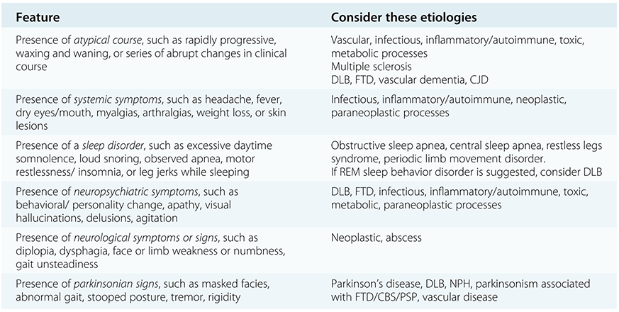
The presentation and course of cognitive decline, and the presence or absence of associated features, are critical elements of the history. Clues suggestive of a non-Alzheimer’s disease etiology of cognitive impairment are listed in Table 1.3. In many instances a seasoned clinician can surmise which etiologic category of disease is likely at play, and a syndromic diagnosis can be made with relative confidence [Reference Knopman, Boeve and Petersen7, Reference Knopman and Petersen8]. For example, an elderly individual with a slowly progressive disorder which has evolved over at least a year, manifesting primarily with anterograde memory impairment with additional features of language and/or visuospatial dysfunction, and relatively normal general physical and neurological examinations, is likely to suffer from a degenerative process with Alzheimer’s disease dementia being the most fitting syndromic diagnosis [Reference McKhann, Knopman and Chertkow9]. Executive and visuospatial impairment along with delusions of misidentification and hallucinations, recurrent dream enactment behavior during sleep suggesting rapid eye movement (REM) sleep behavior disorder, fluctuations in cognition and arousal, and parkinsonism all evolving over one or more years in an older patient suggests dementia with Lewy bodies [Reference McKeith, Dickson and Lowe10]. Changes in behavior/personality and/or language – particularly social disinhibition, loss of empathy, apathy, food preference alterations, – with relative preservation of memory, visuospatial functioning, and praxis suggest behavioral variant frontotemporal dementia [Reference Rascovsky, Hodges and Knopman11]. Such individuals can appear normal in the office setting and on mental status examination, yet their informants (especially when questioned separately) describe behavioral changes that suggest dysfunction in the prefrontal cortex. Vascular dementia is characterized by cognitive changes temporally associated with stroke(s), with clinical and/or radiological evidence of cerebral infarcts [Reference Roman, Tatemichi and Erkinjuntti12].

Table 1.3 Long description
The table has 2 columns: Feature and Consider these etiologies. It reads as follows. Row 1. Feature: Presence of atypical course, such as rapidly progressive, waxing and waning, or series of abrupt changes in clinical course. Etiologies: Vascular, infectious, inflammatory/autoimmune, toxic metabolic processes, Multiple sclerosis, D L B, F T D, vascular dementia, C J D. Row 2. Feature: Presence of systemic symptoms, such as headache, fever, dry eyes/mouth, myalgias, arthralgias, weight loss, or skin lesions. Etiologies: Infectious, inflammatory/autoimmune, neoplastic, paraneoplastic processes. Row 3. Presence of a sleep disorder, such as excessive daytime somnolence, loud snoring, observed apnea, motor restlessness/ insomnia, or leg jerks while sleeping. Etiologies: Obstructive sleep apnea, central sleep apnea, restless legs syndrome, periodic limb movement disorder. If REM sleep behavior disorder is suggested, consider D L B. Row 4. Feature: Presence of neuropsychiatric symptoms, such as behavioral/ personality change, apathy, visual hallucinations, delusions, agitation. Etiologies: D L B, F T D, infectious, inflammatory/autoimmune, toxic, metabolic, paraneoplastic processes. Row 5. Feature: Presence of neurological symptoms or signs, such as diplopia, dysphagia, face or limb weakness or numbness, gait unsteadiness. Etiologies: Neoplastic, abscess. Row 6. Feature: Presence of parkinsonian signs, such as masked facies, abnormal gait, stooped posture, tremor, rigidity. Etiologies: Parkinson's disease, D L B, N P H, parkinsonism associated with F T D/C B S/P S P, vascular disease. The abbreviations are explained at the bottom. C B S: corticobasal syndrome, C J D: Creutzfeldt-Jakob disease, D L B: dementia with Lewy bodies, F T D: frontotemporal dementia, N P H: normal pressure hydrocephalus, P S P: progressive supranuclear palsy.
Abbreviations: CBS = corticobasal syndrome, CJD = Creutzfeldt–Jakob disease, DLB = dementia with Lewy bodies, FTD = frontotemporal dementia, NPH = normal pressure hydrocephalus, PSP = progressive supranuclear palsy.
Cognitive impairment and/or behavioral changes that begins abruptly, markedly fluctuates, or progresses over weeks or months should trigger concerns for a non-degenerative process. In many instances those disorders are potentially treatable. Likewise, symptoms such as headache, fever, skin lesions, significant weight loss, limb paresthesias or weakness, parkinsonism, falls, gait impairment, delusions, visual hallucinations, and incontinence are all features that should raise suspicion of a condition other than Alzheimer’s disease. The presence of depression or suicidality should be queried and these symptoms are particularly prominent in subcortical disorders such as progressive supranuclear palsy, corticobasal degeneration Huntington’s disease and dementia with Lewy bodies. Differentiating pseudodementia (cognitive impairment resulting from depression) from a degenerative dementing illness is often difficult. More often than not, when a person has serious cognitive complaints in conjunction with a mood disorder, treatment of the mood disorder does not fully alleviate the cognitive issues. Still, when depression is strongly suspected, a trial of an antidepressant with few cognitive side effects can provide diagnostic and therapeutic information by virtue of the response to an adequate trial. Often, such a diagnosis and empiric trial has already been carried out without satisfactory results by the time primary care clinicians or patients/family members initiate referrals to dementia specialists. Our task is to determine whether there is an underlying neurodegenerative, infectious, vascular or metabolic condition responsible for the cognitive symptoms.
Investigations
The initial objective of the clinician is to identify and effectively treat all reversible causes or contributors of cognitive impairment. If no such causes or contributors are found, or if the individual continues to experience cognitive impairment once all causes and contributors are addressed, the clinician must then determine (to the extent possible within the context of the patient’s/family’s wishes) which irreversible cause or contributor of impairment is present for management and prognostic purposes. The challenge for the clinician is to determine which diagnostic studies and consultations are necessary for each particular patient who presents with changes in cognition and/or behavior.
Several diagnostic studies available for clinical purposes are shown in Table 1.4. The minimal diagnostic work-up is appropriate for most individuals. A computed tomography (CT) scan of the head with and without contrast is necessary to visualize some tumors and discriminate hemorrhage versus other lesions if they are strongly suspected. A magnetic resonance imaging (MRI) scan of the head offers multiple advantages over CT for demonstrating numerous types of intracranial abnormalities. Contrast enhancement allows visualization of tumors, meningeal processes, and infectious and inflammatory disorders. Usually, contrast is not necessary in the evaluation of a patient for dementia. A standard MRI scan includes sagittal and axial images; coronal images offer additional information as topography and degree of hippocampal and cerebral cortical atrophy are best shown on coronal slices. A sequence that is sensitive to microhemorrhages that are common in Alzheimer’s disease should be performed. When Creutzfeldt–Jakob disease is in question, diffusion-weighted imaging is critical.
Neuropsychological testing
The battery should adequately assess the key cognitive domains
Minimal diagnostic laboratory and imaging battery
Electrolytes, renal/liver function tests: sodium, potassium, calcium, magnesium, creatinine, urea, bilirubin, alkaline phosphatase, liver enzymes
Complete blood count
Thyroid function tests (TSH; often sufficient)
Vitamin B12 and folate
Syphilis serology
Sedimentation rate
Computed tomography (CT) scan of the head or more preferably:
Magnetic resonance imaging (MRI) scan of the head
Additional laboratory studies in select cases
Many to consider based on blood, urine, and cerebrospinal fluid (CSF) – see suggestions in chapters relevant to the syndrome/disorder of interest
Genetic testing
Additional neuroimaging studies in select cases
Perfusion single-photon emission computed tomography (SPECT) (available for clinical use)
Dopamine transporter SPECT imaging (DaTscan) (available for clinical use)
Fluorodeoxyglucose positron emission tomography (FDG-PET) (available for clinical use)
Amyloid-PET (several ligands available for research +/– clinical use)
Tau-PET (ligands available for research)
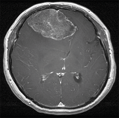
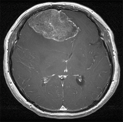
While arguments are sometimes made against the performance of these minimal investigations – particularly for a CT or MRI scan – in every patient with cognitive impairment on the basis of being too costly, the clinician must weigh the costs of these studies for each patient against the potential for identifying a disorder which could impact management and prognosis. Yet not imaging the organ of interest in those with cognitive/behavioral changes can miss important structural etiologies, some of which are treatable – a worthy example is shown in Figure 1.1. Further, patterns of atrophy can be diagnostic. Alzheimer’s disease is associated with hippocampal and posterior temporal parietal atrophy, while the frontotemporal disorders show predominantly frontal atrophy.
Gadolinium-enhanced T1-weighted axial MR image in a 54-year-old woman with an 18-month history of cognitive and behavioral changes who had been treated with sertraline for presumed “pseudodementia” for over 12 months without any improvement in symptoms. A gross total resection of the tumor was performed (histology revealed meningioma) with complete resolution of cognitive/behavioral changes.
The selection of the additional studies and consultations depends on the circumstances of each particular patient. Neuropsychological testing can be very helpful in characterizing the topography and severity of cerebral dysfunction and should be considered in most patients – particularly in those with mild symptoms, and in those who are young or have atypical features.
Blood and cerebrospinal fluid and PET-based biomarkers for Alzheimer’s disease allow the clinician to rule in a diagnosis of this condition and increasingly are becoming the standard for diagnosis. Similarly, real-time quaking-induced conversion (RT-QuIC) tests for alpha-synuclein are increasingly more sensitive for picking up Lewy body disease pathology.
Management
Basic Principles
Several strategies in the management of patients with dementia are shown in Table 1.5.
Ensure infections, sleep disorders, dehydration, metabolic disturbances, pain, and constipation are being effectively treated
Correct hearing or visual loss to the extent possible
Avoid agents with anticholinergic properties
Minimize psychoactive medications with possible adverse cognitive side effects to the fewest agents at the lowest effective doses
Promote regular sleep–wake times and daily routine
Regular light exercise
Caregiver support – support groups, respite care, etc.
Consider participating in research, clinical trials
Pharmacological
Cognition and behavior reflect the integrated coordination of neural networks within the brain. When one or more of these networks are affected by an abnormal process and neurochemical systems are altered, pharmacotherapy (i.e., “symptomatic therapies”) directed at these alterations can improve symptoms. These symptomatic therapies are discussed in the pertinent chapters. Antibodies directed against the amyloid protein have been proven to positively influence dementia-associated disease mechanisms in humans and must be considered in patients with Alzheimer’s disease (AD). Disease-modifying therapies continue for AD and are beginning with FTD-spectrum disorders and the parkinsonian-dementia syndromes.
Information
All families facing dementia-related issues should be informed about national and international organizations devoted to dementia education and support. An abbreviated list of key organizations is shown in Table 1.6.
Alzheimer’s Disease
Alzheimer’s Association
Alzheimer Society of Canada
Alzheimer Society of the United Kingdom
Alzheimer’s Australia Dementia Research Foundation
National Institute on Aging – Information on AD
Dementia with Lewy Bodies/Lewy Body Dementia
Lewy Body Dementia Association
Lewy Body Society
National Institute on Aging – Information on DLB
www.nia.nih.gov/alzheimers/publication/lewy-body-dementia/introduction
Frontotemporal Dementia/Frontotemporal Degeneration
Association for Frontotemporal Degeneration
National Institute on Aging – Information on FTD
www.nia.nih.gov/alzheimers/publication/frontotemporal-disorders/basics-frontotemporal-disorders
Normal Pressure Hydrocephalus
Normal Pressure Hydrocephalus
Creutzfeldt–Jakob Disease
Creutzfeldt–Jakob Disease Foundation
* This is only a partial list; numerous other reputable websites pertinent to dementia-related issues can be found on the internet.
Several institutions conduct longitudinal studies on aging and dementia and/or conduct experimental drug trials, and the Alzheimer’s Association can facilitate identifying a nearby institution involved in such studies. The ClinicalTrial.gov website is also reputable and updated frequently: www.clinicaltrials.gov.
It should be mentioned that some information on the internet is obviously inaccurate, and patients and families should be encouraged to access information from reputable sources and discuss questionable information with their physicians.