




No CrossRef data available.
Article contents
The sleeping brain and the neural basis of emotions
Published online by Cambridge University Press: 23 May 2012
Abstract
In addition to active wake, emotions are generated and experienced in a variety of functionally different states such as those of sleep, during which external stimulation and cognitive control are lacking. The neural basis of emotions can be specified by regarding the multitude of emotion-related brain states, as well as the distinct neuro- and psychodynamic stages (generation and regulation) of emotional experience.
Information
- Type
- Open Peer Commentary
- Information
- Copyright
- Copyright © Cambridge University Press 2012
References
Benca, R. M., Obermeyer, W. H., Thisted, R. A. & Gillin, J. C. (1992) Sleep and psychiatric disorders. A meta-analysis. Archives of General Psychiatry
49:651–68; discussion 669–70 .CrossRefGoogle ScholarPubMed
Braun, A. R., Balkin, T. J., Wesenten, N. J., Carson, R. E., Varga, M., Baldwin, P., Selbie, S., Belenky, G. & Herscovitch, P. (1997) Regional cerebral blood flow throughout the sleep-wake cycle. An H2(15)O PET study. Brain
120:1173–97.CrossRefGoogle ScholarPubMed
Braun, A. R., Balkin, T. J., Wesensten, N. J., Gwadry, F., Carson, R. E., Varga, M., Baldwin, P., Belenky, G. & Herscovitch, P. (1998) Dissociated pattern of activity in visual cortices and their projections during human rapid eye movement sleep. Science
279:91–95.CrossRefGoogle ScholarPubMed
Fosse, R., Stickgold, R. & Hobson, J. A. (2004) Thinking and hallucinating: Reciprocal changes in sleep. Psychophysiology
41:298–305.CrossRefGoogle ScholarPubMed
Foulkes, W. D. (1962) Dream reports from different stages of sleep. Journal of Abnormal and Social Psychology
65:14–25.CrossRefGoogle ScholarPubMed
Fuster, J. M. (2006) The cognit: A network model of cortical representation. International Journal of Psychophysiology
60:125–32. Available at: http://www.ncbi.nlm.nih.gov/pubmed/16626831.CrossRefGoogle ScholarPubMed
Gastaut, H. & Broughton, R. (1964) A clinical and polygraphic study of episodic phenomena during sleep. Recent Advances in Biological Psychiatry
7:197–221.Google Scholar
Gottesmann, C. (1999) Neurophysiological support of consciousness during waking and sleep. Progress in Neurobiology
59:469–508.CrossRefGoogle ScholarPubMed
Gottesmann, C. (2010) The development of the science of dreaming. International Review of Neurobiology
92:1–29.CrossRefGoogle ScholarPubMed
Hobson, J. A. (1999) Dreaming as a delirium: How the brain does out of its mind. MIT Press.CrossRefGoogle Scholar
Hobson, J. A. (2009) REM sleep and dreaming: Towards a theory of protoconsciousness. Nature Reviews Neuroscience
10:803–13.CrossRefGoogle ScholarPubMed
Hobson, J. A. & Pace-Schott, E. F. (2002) The cognitive neuroscience of sleep: Neuronal systems, consciousness and learning. Nature Reviews Neuroscience
3:679–93.CrossRefGoogle ScholarPubMed
Hobson, J. A., Pace-Schott, E. F. & Stickgold, R. (2000) Dreaming and the brain: Toward a cognitive neuroscience of conscious states. Behavioral and Brain Sciences
23:793–842; discussion 904–1121.CrossRefGoogle Scholar
Maquet, P., Péters, J., Aerts, J., Delfiore, G., Degueldre, C., Luxen, A. & Franck, G. (1996) Functional neuroanatomy of human rapid-eye-movement sleep and dreaming. Nature
383:163–66.CrossRefGoogle ScholarPubMed
McNamara, P., Johnson, P., McLaren, D., Harris, E., Beauharnais, C. & Auerbach, S. (2010) REM and NREM sleep mentation. International Review of Neurobiology
92:69–86.CrossRefGoogle ScholarPubMed
Morris, J. S., Ohman, A. & Dolan, R. J. (1998) Conscious and unconscious emotional learning in the human amygdala. Nature
393:467–70.CrossRefGoogle ScholarPubMed
Morris, J. S., Ohman, A. & Dolan, R. J. (1999) A subcortical pathway to the right amygdala mediating “unseen” fear. Proceedings of the National Academy of Sciences USA
96:1680–85.CrossRefGoogle Scholar
Nofzinger, E. A., Mintun, M. A., Wiseman, M., Kupfer, D. J. & Moore, R. Y. (1997) Forebrain activation in REM sleep: An FDG PET study. Brain Research
770:192–201.CrossRefGoogle ScholarPubMed
Phillips, M. L., Drevets, W. C., Rauch, S. L. & Lane, R. (2003) Neurobiology of emotion perception I: The neural basis of normal emotion perception. Biological Psychiatry
54:504–14.CrossRefGoogle ScholarPubMed
Stickgold, R., Hobson, J. A., Fosse, R. & Fosse, M. (2001) Sleep, learning, and dreams: Off-line memory reprocessing. Science
294:1052–57.CrossRefGoogle ScholarPubMed
Tamietto, M. & de Gelder, B. (2010) Neural bases of the non-conscious perception of emotional signals. Nature Reviews Neuroscience
11:697–709.CrossRefGoogle ScholarPubMed
Tononi, G. & Koch, C. (2008) The neural correlates of consciousness: An update. Annals of the New York Academy of Sciences
1124:239–61.CrossRefGoogle ScholarPubMed
Van der Helm, E., Gujar, N. & Walker, M. P. (2010) Sleep deprivation impairs the accurate recognition of human emotions. Sleep
33:335–42.CrossRefGoogle ScholarPubMed
Vuilleumier, P., Armony, J. L., Driver, J. & Dolan, R. J. (2001) Effects of attention and emotion on face processing in the human brain: An event-related fMRI study. Neuron
30:829–41.CrossRefGoogle Scholar
Walker, M. P. (2009) The role of sleep in cognition and emotion. Annals of the New York Academy of Sciences
1156:168–97.CrossRefGoogle ScholarPubMed
Walker, M. P. & van der Helm, E. (2009) Overnight therapy? The role of sleep in emotional brain processing. Psychological Bulletin
135:731–48.CrossRefGoogle ScholarPubMed
Williams, L. M., Das, P., Liddell, B. J., Kemp, A. H., Rennie, C. J. & Gordon, E. (2006) Mode of functional connectivity in amygdala pathways dissociates level of awareness for signals of fear. Journal of Neuroscience
26:9264–71.CrossRefGoogle ScholarPubMed
Yoo, S. S., Gujar, N., Hu, P., Jolesz, F. A. & Walker, M. P. (2007) The human emotional brain without sleep – a prefrontal amygdala disconnect. Current Biology
17:R877–78.CrossRefGoogle ScholarPubMed
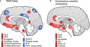
Figure 1. Regional activation patterns: (A) Blue (white numbers) and red (black numbers) areas indicate regions showing deactivation and hyperactivation, respectively, during REM sleep compared to wake (modification of Hobson 2009). (B) System of non-conscious emotion processing (modification of Tamietto & de Gelder 2010). ACC: anterior cingulate cortex; OFC: orbitofrontal cortex; Parahipp.: parahippocampal; DLPFC: dorsolateral prefrontal cortices; PCC: posterior cingulate cortex; Prim.: primary; NA: nucleus accumbens; PAG: periaqueductal gray; LC: locus coeruleus. A color version of this image can be viewed at http://www.journals.cambridge.org/bbs.
According to the “conceptual act model,” emotional experience is a result of a complex psychological operation termed “situated conceptualization,” which gives a meaningful context to a basic core affect. Conceptualization is guided by fundamental cognitive processes such as retrieval of stored memory representations and executive attention. Accordingly, the neural basis of emotion processing is stated to be represented by a variety of interacting brain regions, which are traditionally associated with both basic psychological operations and emotional experience. In substantiating the role of conceptualization, the model accounts for the active states of the waking human brain, during which goal-directed behaviors induce a relevant context.
Critically, emotion categories can be generated during brain states which essentially differ from active wake in that goal-directed executive control networks are not active. Neuroimaging studies have demonstrated that brain areas including the dorsolateral prefrontal cortices (DLPFC), parietal cortices, precuneus, posterior cingulate cortex (PCC), and primary visual cortex, all of which support higher cognitive functions and executive control (Fuster Reference Fuster2006), are suppressed during rapid eye movement (REM) sleep compared with wake; whereas the anterior cingulate cortex (ACC), orbitofrontal cortex (OFC), amygdaloid complexes, pontine tegmentum, parahippocampal cortex, and extrastriate visual cortices are more active in REM sleep than in wake and non-REM sleep (Braun et al. Reference Braun, Balkin, Wesenten, Carson, Varga, Baldwin, Selbie, Belenky and Herscovitch1997; Reference Braun, Balkin, Wesensten, Gwadry, Carson, Varga, Baldwin, Belenky and Herscovitch1998; Maquet et al. Reference Maquet, Péters, Aerts, Delfiore, Degueldre, Luxen and Franck1996; Nofzinger et al. Reference Nofzinger, Mintun, Wiseman, Kupfer and Moore1997) (see our Figure 1a). Yet, intensive emotional experience does exist during REM sleep, and the emotional categories of REM sleep do not differ from those experienced during wake (Fosse et al. Reference Fosse, Stickgold and Hobson2004; Hobson et al. Reference Hobson, Pace-Schott and Stickgold2000; McNamara et al. Reference McNamara, Johnson, McLaren, Harris, Beauharnais and Auerbach2010; Stickgold et al. Reference Stickgold, Hobson, Fosse and Fosse2001; Walker & van der Helm Reference Walker and van der Helm2009). Importantly, in REM sleep, emotions emerge out of goal-directed behavioral context and under a lack of external input. Also, their conceptualization and goal-directed reference are elusive (Fosse et al. Reference Fosse, Stickgold and Hobson2004; Hobson Reference Hobson1999; Reference Hobson2009; Hobson et al. Reference Hobson, Pace-Schott and Stickgold2000) as implied by the strong DLFC suppression during REM sleep.
Regional activation patterns: (A) Blue (white numbers) and red (black numbers) areas indicate regions showing deactivation and hyperactivation, respectively, during REM sleep compared to wake (modification of Hobson 2009). (B) System of non-conscious emotion processing (modification of Tamietto & de Gelder 2010). ACC: anterior cingulate cortex; OFC: orbitofrontal cortex; Parahipp.: parahippocampal; DLPFC: dorsolateral prefrontal cortices; PCC: posterior cingulate cortex; Prim.: primary; NA: nucleus accumbens; PAG: periaqueductal gray; LC: locus coeruleus. A color version of this image can be viewed at http://www.journals.cambridge.org/bbs.
In non-REM sleep, and particularly in slow wave sleep (SWS), the activity of all these brain areas is suppressed (Braun et al. Reference Braun, Balkin, Wesenten, Carson, Varga, Baldwin, Selbie, Belenky and Herscovitch1997; Maquet et al. Reference Maquet, Péters, Aerts, Delfiore, Degueldre, Luxen and Franck1996; Nofzinger et al. Reference Nofzinger, Mintun, Wiseman, Kupfer and Moore1997) and mental experience is extremely scarce (Hobson & Pace-Schott Reference Hobson and Pace-Schott2002). Nonetheless, although reports from non-REM sleep are often thought-like (Hobson Reference Hobson1999; Hobson et al. Reference Hobson, Pace-Schott and Stickgold2000), signs of emotions reflecting an individual's current concerns can be detected (Foulkes Reference Foulkes1962; Hobson et al. Reference Hobson, Pace-Schott and Stickgold2000). Moreover, night terrors (pavor nocturnos) characterized by a general feeling of fear accompanied by intense autonomic discharge, typically occur during the deepest stage 4 of SWS (Gastaut & Broughton Reference Gastaut and Broughton1964; Gottesmann Reference Gottesmann2010). Thus, substantially different brain states such as those of wake, and non-REM and REM sleep – characterized also by dramatic differences in mentality, neurochemistry, connectivity, and neuroelectric signaling (Gottesmann Reference Gottesmann1999; Hobson & Pace-Schott Reference Hobson and Pace-Schott2002; Hobson et al. Reference Hobson, Pace-Schott and Stickgold2000; Stickgold et al. Reference Stickgold, Hobson, Fosse and Fosse2001; Tononi & Koch Reference Tononi and Koch2008) – can all provide a neural basis for the experience of distinct emotional categories. Further, in a variety of dysfunctional wake states associated with psychopathological and neurological deficits, sleep alterations (e.g., Benca et al. Reference Benca, Obermeyer, Thisted and Gillin1992) or sleep deprivation (Van der Helm et al. Reference Van der Helm, Gujar and Walker2010; Yoo et al. Reference Yoo, Gujar, Hu, Jolesz and Walker2007), in which critical brain regions and/or their connections are functionally altered (over-activated or de-activated), emotion generation is preserved, although subjective emotional experience can be modulated (Walker Reference Walker2009).
State-related, in particular sleep-related, emotions can be viewed in the context of two existing concepts regarding the distinction between conscious and non-conscious processing of emotions, and the neurodynamics of emotional perception: (1) Increasing evidence is provided for the coexistence of two distinct neural systems in the waking brain, which subserve conscious and non-conscious processing of emotions (Morris et al. Reference Morris, Ohman and Dolan1998; Vuilleumier et al. Reference Vuilleumier, Armony, Driver and Dolan2001). Non-conscious perception of emotional stimuli has been associated with the functional integrity of a subcortical network including the pulvinar, amygdala, nucleus accumbens, periaqueductal gray, and locus coeruleus (Morris et al. Reference Morris, Ohman and Dolan1999; Tamietto & de Gelder Reference Tamietto and de Gelder2010; Williams et al. Reference Williams, Das, Liddell, Kemp, Rennie and Gordon2006) (see our Figure 1b), which may function independently of cortical areas. It is proposed that this network incorporating also the ACC and OFC (Fig. 1b) is genetically established and phylogenetically adaptive, and can, through rapid feed-forward influences, enhance the pre-attentive processing of emotional signals during goal-directed behavior. It is only during wake that an executive cortical feedback is suggested to exert inhibitory modulation over emotion-related subcortical areas (Tamietto & de Gelder Reference Tamietto and de Gelder2010). It is notable that major components of the non-conscious emotional system are overactivated during REM sleep relative to wake (ACC, OFC, and amygdala – see Fig. 1) implying enhanced emotion processing within this system due to the lack of inhibitory modulation from cognitive cortical areas. (2) Emotion perception is proposed to be underlain by three neurophysiological stages: (i) identification of emotional significance of information; (ii) production of affective state including the generation of autonomic, neuroendocrine, and neuromuscular responses and subjective responses of emotional experience; and (iii) regulation of the affective state (Phillips et al. Reference Phillips, Drevets, Rauch and Lane2003).
Within these concepts, physiological and subjective experience markers indicate that affective states are generated during sleep (Hobson & Pace-Schott Reference Hobson and Pace-Schott2002), but they are not regulated by cortical regions (Fig. 1). Emotion generation during sleep is entirely driven by internal information, being unaffected by operations subserving the interaction with the external environment (Hobson Reference Hobson1999). Thus, the emerging emotional categories may not be an online integrative product of ongoing context appraisal (as Lindquist et al. suggest in the target article). Rather, emotional dream experience reflects the functioning of local (Tononi & Koch Reference Tononi and Koch2008) or primary consciousness (protoconsciousness) defined as simple awareness of perception and emotions (Hobson Reference Hobson2009).
We argue that emotions emerge in a variety of distinct brain states. The neural mechanisms of emotion generation are different from the neural mechanisms of emotional regulation, with the latter being able to modulate emotion production only during conscious processing. We suggest that the neural substrate of emotional categories can be identified adequately by exploring different functional brain states, in which emotion generation may not be dominated by the executive control mechanisms. Thus, models of the brain basis of emotion would demarcate the neural substrates of emotion generation, emotional experience, and emotional regulation.