




No CrossRef data available.
Article contents
A rapprochement between emotion and cognition: Amygdala, emotion, and self-relevance in episodic-autobiographical memory
Published online by Cambridge University Press: 23 May 2012
Abstract
Lindquist et al. remark that not all fear instances lead to heightened amygdalar activity and, instead, point to roles of the amygdala in detecting “motivationally salient “or “emotionally impactful” stimuli. By reviewing research on the amygdala's functions in episodic-autobiographical memory, we further emphasize the involvement of the amygdala in coding the subjective relevance and extracting the biological and social significance of the stimuli.
Information
- Type
- Open Peer Commentary
- Information
- Copyright
- Copyright © Cambridge University Press 2012
References
Bartlett, F. & John, E. R. (1973) Equipotentiality quantified: The anatomical distribution of the engram. Science
181:764–67.CrossRefGoogle ScholarPubMed
Chow, K. L. (1967) Effects of ablation. In: The neurosciences, ed. Quarton, G. C., Melnechuk, T. & Schmitt, F. O., pp. 705–13. Rockefeller University Press.Google Scholar
Daselaar, S. M., Rice, H. J., Greenberg, D. L., Cabeza, R., LaBar, K. S. & Rubin, D. C. (2008) The spatiotemporal dynamics of autobiographical memory: Neural correlates of recall, emotional intensity, and reliving. Cerebral Cortex
18:217–29.CrossRefGoogle ScholarPubMed
Eich, E., Nelson, A. L., Leghari, M. D. & Handy, T. C. (2009) Neural systems mediating field and observer memories. Neuropsychologia
47:2239–51.CrossRefGoogle ScholarPubMed
Fink, G. R., Markowitsch, H. J., Reinkemeier, M., Bruckbauer, T., Kessler, J. & Heiss, W.-D. (1996) Cerebral representation of one's own past: Neural networks involved in autobiographical memory. Journal of Neuroscience
16:4275–82.CrossRefGoogle ScholarPubMed
John, E. R. (1972) Switchboard versus statistical theories of learning and memory. Science
177:850–64.CrossRefGoogle ScholarPubMed
Kanwisher, N. (2010) Functional specificity in the human brain: A window into the functional architecture of the mind. Proceedings of the National Academy of Sciences USA
107(25):11163–70.CrossRefGoogle Scholar
Markowitsch, H. J. (1994) The memory storehouse. Trends in Neuroscience
17:513 (Letter).CrossRefGoogle ScholarPubMed
Markowitsch, H. J. (1999) Limbic system. In: The MIT encyclopedia of the cognitive sciences, ed. Wilson, R. & Keil, F., pp. 472–75. MIT Press.Google Scholar
Markowitsch, H. J., Calabrese, P., Würker, M., Durwen, H. F., Kessler, J., Babinsky, R., Brechtelsbauer, D., Heuser, L. & Gehlen, W. (1994) The amygdala's contribution to memory – A PET-study on two patients with Urbach-Wiethe disease. NeuroReport
5:1349–52.Google Scholar
Markowitsch, H. J. & Staniloiu, A. (2011) Amygdala in action: Relaying biological and social significance to autobiographic memory. Neuropsychologia
49:718–33.CrossRefGoogle Scholar
Markowitsch, H. J., Thiel, A., Reinkemeier, M., Kessler, J., Koyuncu, A. & Heiss, W.-D. (2000) Right amygdalar and temporofrontal activation during autobiographic, but not during fictitious memory retrieval. Behavioural Neurology
12:181–90.CrossRefGoogle Scholar
Papez, J. W. (1937) A proposed mechanism of emotion. Archives of Neurology and Psychiatry
38:725–43.CrossRefGoogle Scholar
Schulte-Rüther, M., Greimell, E., Markowitsch, H. J., Kamp-Becker, I., Remschmidt, H., Fink, G. R. & Piefke, M. (2010) Dysfunctional brain networks supporting empathy in adults with autism spectrum disorder: An fMRI study. Social Neuroscience
6:1–21.CrossRefGoogle Scholar
Sharot, T., Martorella, E. A., Delgado, M. R. & Phelps, E. A. (2007) How personal experience modulates the neural circuitry of memories of September 11. Proceedings of the National Academy of Sciences USA
104:389–94.CrossRefGoogle Scholar
Siebert, M., Markowitsch, H. J. & Bartel, P. (2003) Amygdala, affect, and cognition: Evidence from ten patients with Urbach-Wiethe disease. Brain
126:2627–37.CrossRefGoogle ScholarPubMed
Staniloiu, A. & Markowitsch, H. J. (2010) Looking at comorbidity through the glasses of neuroscientific memory research: A brain network perspective. Behavioral and Brain Sciences
33:171–72.CrossRefGoogle ScholarPubMed
von Cramon, D. Y. & Markowitsch, H. J. (1992) The problem of “localizing” memory in focal cerebro-vascular lesions. In: Neuropsychology of memory, 2nd edition, ed. Squire, L. R. & Butters, N., pp. 95–105. Guilford Press.Google Scholar
von Cramon, D. Y. & Markowitsch, H. J. (2000) The septum and human memory. In: The behavioral neuroscience of the septal region, ed. Numan, R., pp. 380–413. Springer.CrossRefGoogle Scholar
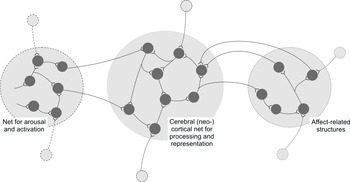
Figure 1. A schematic sketch of a neural net composed of three interacting subsets.
As Lindquist et al. describe, oscillations between localization and anti-localization of functions characterize brain research. Localizationists argue that circumscribed brain lesions can abolish a function throughout a life-span. Frequently cited is the patient H.M. who, after undergoing bilateral resection of the medial temporal lobes, developed life-long anterograde amnesia (Markowitsch & Staniloiu, in press). Functional neuroimaging data also point to specialized regions being selectively activated during face processing (Kanwisher Reference Kanwisher2010). At the other pole of the dialectics, Chow (Reference Chow, Quarton, Melnechuk and Schmitt1967) argued that if a lesion in a brain region has no particular effect on a function, it cannot be concluded that this region is irrelevant for that function in non-damaged individuals. Second, if a lesion does influence the performance of a task, this does not mean that that region is the only structure involved in the task. And last, and perhaps most important, brain tissue damage cannot clarify the function of a brain area, because it destroys what it aims to study – that brain area. Functional imaging studies, showing that widespread regions in the brain are activated even when performed tasks are simple, and electrophysiological experiments (Bartlett & John Reference Bartlett and John1973; John Reference John1972) support views of the brain acting in a Gestalt-like fashion.
Generally, no brain region can act alone in vivo, and, especially for more complex functions, a multitude of brain regions act in concert or within a neural network (Cramon & Markowitsch Reference von Cramon, Markowitsch, Squire and Butters1992; Fink et al. Reference Fink, Markowitsch, Reinkemeier, Bruckbauer, Kessler and Heiss1996; Markowitsch Reference Markowitsch1994; Schulte-Rüther et al. Reference Schulte-Rüther, Greimell, Markowitsch, Kamp-Becker, Remschmidt, Fink and Piefke2010; Staniloiu & Markowitsch Reference Staniloiu and Markowitsch2010). This was already suggested by Papez in 1937 in his seminal paper entitled “A proposed mechanism of emotion” (Papez [Reference Papez1937], overlooked by the Lindquist et al. in the target article). Processing of episodic-autobiographical memories requires attention and concentration, self-reflection and emotional colorization (Markowitsch & Staniloiu, in press) (see our Fig. 1).
A schematic sketch of a neural net composed of three interacting subsets.
Emotional evaluation and tagging are important for both formation and retrieval of episodic-autobiographical memories, and they engage limbic system regions (Markowitsch Reference Markowitsch, Wilson and Keil1999). Within these regions, the amygdala constitutes a cornerstone as it represents the major hub for channeling sensory information of biological or social personal significance to storage in networks located primarily in neocortical areas. Behavioral studies performed in patients with Urbach-Wiethe disease, a rare genetic condition leading to bilateral calcification of the amygdala (Cahill et al. 1995; Markowitsch et al. Reference Markowitsch, Calabrese, Würker, Durwen, Kessler, Babinsky, Brechtelsbauer, Heuser and Gehlen1994) emphasize the role of the amygdala for evaluating the salience of newly incoming information and extracting those portions which appear worth being remembered.
Several combined neuroimaging and behavioral studies of healthy subjects or patients showed amygdalar engagement in episodic-autobiographical mnemonic processing. Brain mapping of the time course of episodic-autobiographical memory retrieval revealed modulation of amygdala activity by judgments of emotional intensity of autobiographical memory. This occurred early in the process of retrieval, before (healthy) participants in the study signaled the full assemblage of autobiographical memories (Daselaar et al. Reference Daselaar, Rice, Greenberg, Cabeza, LaBar and Rubin2008). This finding is congruent with the proposed role of the amygdala in charging sensory information with appropriate emotional cues, in order to guide successful searching for and retrieval of emotionally significant events (Markowitsch & Staniloiu Reference Markowitsch and Staniloiu2011).
Not all functional neuroimaging investigations of episodic-autobiographical memory retrieval evidenced amygdalar activation, due to differences in memory-testing methodology, imaging techniques' limitations, lack of control for retrieval perspective, or habituation of amygdala responses to emotional material over time (Markowitsch & Staniloiu Reference Markowitsch and Staniloiu2011). An increase in right amygdalar activity was found in subjects who retrieved episodic-autobiographical memories from a first person perspective, in comparison to those who retrieved them from a third person perspective (Eich et al. Reference Eich, Nelson, Leghari and Handy2009), suggesting a higher degree of subjectively perceived emotionality associated with the first type of memories. Neuroimaging studies showed activation of the amygdala as a function of personal relevance or self-involvement of the recalled material. One study comparing the retrieval of autobiographical episodes with the retrieval of fictitious “episodes” revealed activation of the amygdala only during the recall of personal – that is, authentic – events, whereas the recall of fictitious material activated the retrosplenial/precuneus area only (Markowitsch et al. Reference Markowitsch, Thiel, Reinkemeier, Kessler, Koyuncu and Heiss2000). Personal relevance enhanced amygdala activity during the recall of the frightening event of September 11, 2001 (Sharot et al. Reference Sharot, Martorella, Delgado and Phelps2007).
These results suggest that the amygdala has functions in coding the subjective relevance and social and biological salience of the stimuli, which might be reflected in its structure and hodology. The amygdaloid nuclei are heavily connected with subcortical and cortical areas, including regions involved in self-referential processing, such as the ventro-medial prefrontal cortex. From the neocortex, fibers arising from the multisensory posterior association cortex, but in part also from primary sensory regions (olfaction), project to the amygdala. Subsequently, the amygdala receives largely preprocessed sensory input, which then provides a basis for the amgdala to extract the personal biological and social significance of the environment. Nevertheless, the amygdala is not the only structure which plays a role in this process of evaluation. A working tandem may, for example, exist between the amygdala and septal nuclei. A damaged amygdala dampens emotional evaluation (Siebert et al. Reference Siebert, Markowitsch and Bartel2003) whereas damaged septal nuclei enhance it (Cramon & Markowitsch Reference von Cramon, Markowitsch and Numan2000). A patient with septal damage, for example, could not tolerate listening to an emotional story with negatively valenced content, because of overwhelming perceived emotionality (Cramon et al. 1993).
In conclusion, our findings from patients with Urbach-Wiethe disease and from studying episodic-autobiographical memory with various functional imaging techniques in different populations, support Lindquist et al.'s view that the amygdala is not simply an emotional brain structure, but integrates emotion with cognition. This view was, in our case, significantly shaped by work with patients with relatively selective amygdala lesions. This work enabled verifying hypotheses, which may not have been possible by simply resorting to data from imaging studies. Despite using various mathematical models to interpret functional magnetic resonance imaging data, it is still difficult to infer causality from function–structure correlations obtained with brain imaging; therefore, additional experimental techniques (e.g., transcranial magnetic stimulation) may need to be used to confirm whether a brain region is necessary or specific for a given function.