


Highlights
-
Endoscopic endonasal approach is effective as well as safe for anterior skull base meningiomas.
-
This approach showed a low recurrence rate.
-
The main challenge with this procedure is the risk of CSF leak.
Introduction
Anterior skull base meningiomas are a heterogeneous group of tumors classified based on their primary site of attachment. Reference McDermott, Wilson and Youmans1 They account for approximately 6–13% of all meningiomas and exhibit variable clinical presentations. Reference Solero, Giombini and Morello2 These lesions present unique surgical challenges due to their proximity to critical neurovascular structures.
Surgical intervention may be considered depending on the tumor’s size, anatomical relationships and associated neurological deficits. Reference Woodworth3 Among the various surgical approaches, the endoscopic endonasal approach (EEA) offers several advantages, including early devascularization of the lesion, adequate tumor resection and preservation of neurovascular structures. Reference de Divitiis, Cappabianca and Cavallo4,Reference Kassam, Carrau, Snyderman, Gardner and Mintz5 Additionally, EEA enables complete resection of the tumor’s site of attachment while minimizing brain retraction, potentially leading to improved long-term outcomes for patients. Reference Zoli, Guaraldi, Pasquini, Frank and Mazzatenta6,Reference Kshettry, Elshazly and Evans7
This study aims to evaluate the clinical outcomes of patients with anterior skull base meningiomas who underwent EEA for tumor resection. It analyzes clinical presentation, extent of resection, complications, recurrence patterns and prognostic factors. These findings will help facilitate individualized preoperative discussions, guide surgical technique selection and serve as benchmarks for surgeons utilizing this approach.
Methods
The Ottawa Hospital Civic Campus skull base database was queried to identify cases of endoscopic endonasal skull base surgery for the resection of anterior skull base meningiomas between October 2014 and October 2023. Records of all adult patients with this type of lesion were screened for inclusión. Exclusion criteria included the inability to confirm tumor pathology, incomplete clinical records and patients with a history of previous CSF leaks, skull base trauma or active infection at the time of surgery.
All patient medical records and operative notes were reviewed in detail. Data collected included the following: ASA class, type of approach, tumor location and volume (Using the modified ellipsoid formula (A*B*C)/2), surgical time, blood loss, length of stay (LOS), use of lumbar drain, use of dural sealants, use of nasoseptal flap, CSF leak presence, intra operative complications, readmissions at 30 days, World Health Organization (WHO) pathological tumor grade, extent of resection, mean follow up and recurrence at last follow up. Visual evoked potential monitoring was employed in all cases to guide surgical maneuvers and minimize the risk of optic apparatus injury.
Surgical approaches were categorized based on tumor location and site of primary attachments between olfactory groove and tubercullum sellae-planum sphenoidale areas. The olfactory grooves (OGs) of the cribriform plate are shallow depressions located within the ethmoid bone in the anterior cranial fossa that contain the olfactory bulb of the olfactory nerve. The floor is formed by the medial lamella of the cribriform plate, bordered laterally by the lateral lamella and medially by the perpendicular plate. Reference Joiner and van der Kogel8 The tuberculum sellae (TS) is the ridged process of the sphenoid bone, which forms the anterior wall of the sella turcica, and anteriorly, it joins the planum sphenoidale, which is related to the anterior horizontal part of the superior surface of the body of the sphenoid bone Reference Chmielewski9 (Figure 1).
Sagittal and axial reconstructions of the skull base. In purple is the olfactory groove region, and in pink is the planum sphenoidale-tubercullum sellae region.

Open and endoscopic resection approaches were discussed in multidisciplinary skull base rounds, taking into account tumor extension, neurovascular relationships, olfactory function and patient preference. The endoscopic instruments and techniques required to access this areas have been discussed extensively Reference Kassam, Carrau, Snyderman, Gardner and Mintz5,Reference Dehdashti, Ganna, Witterick and Gentili10,Reference Har-El and Casiano11 (Figures 2 and 3). Lumbar drain was used pre emptively in all cases. After satisfactory resection was achieved, skull base reconstruction was performed using an underlay dural substitute, a fascia lata graft and an overlying nasoseptal flap. The construct was further reinforced with fibrin glue and Gelfoam.
Sagittal and axial MRI (Top) depicting a planum-tubercullum sellae meningioma. Endoscopic endonasal approach achieved complete resection of the lesion (Bottom).

Sagittal and axial MRI (Top) depicting an olfactory groove meningioma. Endoscopic endonasal approach achieved complete resection of the lesion (Bottom).

The extent of resection was classified as total, near total (superior to 95% of the tumor) and subtotal. The goal of surgery was gross total resection (GTR) of the tumor in all but four cases with giant or multicompartimental lesions. The degree of tumor resection was evaluated on postoperative imaging studies and included MRI in all cases. The follow up of the patients included 3T brain MRI imaging in order to assess residual/recurrent tumoral tissue.
Descriptive statistics are presented as mean, standard deviation and range for continuous variables, and as frequency and percentage for categorical variables. The Shapiro–Wilk test was applied to ensure normal distribution of the data. The Chi-square test, along with Fisher’s exact test, was used to compare categorical variables and determine relationships between them, with a 95% significance level. Statistical analysis was performed using IBM SPSS Statistics version 26.0 software.
Results
Twenty-five patients underwent an extended EEA for the resection of anterior skull base meningiomas. Seventeen patients (68%) had meningiomas located in the TS–planum sphenoidale region, while eight patients (32%) had OG meningiomas.
The mean preoperative tumor volume was 7.04 cm3 (SD 5.04) in the TS group and 19.54 cm3 (SD 16.4) in the OG group. This difference in tumor volume was statistically significant (p = 0.03). Preoperative ASA classification was type III in 21 patients (84%) and type IV in the remaining four patients (16%) (Table 1).
Patient demographics and general characteristics

The clinical presentation varied depending on the primary tumor location. In the OG group, the most common symptoms were headaches and hyposmia, whereas visual disturbances were the most frequently reported complaints in the TS group. One TS patient presented with third nerve palsy due to a multicompartmental meningioma with cavernous sinus extension.
The mean surgical duration for the endoscopic procedures was 351 minutes (range: 141–711 min, SD 128 min). A preoperative lumbar drain was placed in 40% of cases (2 OG, 8 TS; p = 0.294), and dural sealants were used in 14 patients (5 OG, 9 TS; p = 0.65). Skull base reconstruction involved the use of a nasoseptal flap in 92% of cases. The flap was not utilized in two TS cases (8%).
The mean blood loss was 472 mL (range: 100–2000 mL, SD 431 mL). A major vascular injury occurred in one TS case where the right posterior communicating artery was encased by the tumor. This factor was evident pre operatively. No ischemic complications resulted from this event. Red blood cell transfusion was required in three cases (12%), while fresh frozen plasma transfusion was necessary in two cases (8%). No cases were aborted, and no perioperative mortality was documented. The mean LOS was 11.1 days (range: 2–32 days, SD 6.81). In one case, prolonged hospitalization was required due to a medical complication unrelated to the surgical intervention.
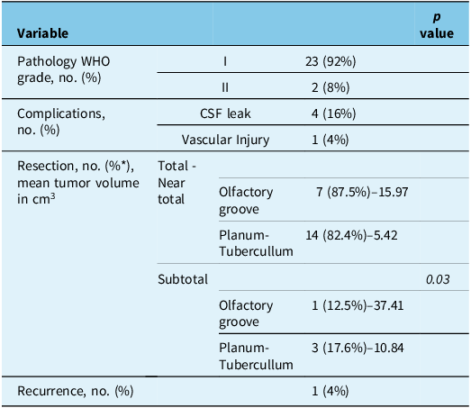
CSF leaks were documented in four cases, all in the TS group. Two patients required readmission within 30 days postoperatively for CSF leak management. All cases were successfully treated with lumbar drain placement. None of these patients required reoperation, and no postoperative infections were reported (Table 2).
Post operative characteristics

* Based on tumor location.
Total or near-total resection was achieved in 87.5% of OG cases and 82.4% of TS cases. Subtotal tumor resection was performed in four cases: One patient in the OG group with a giant OG meningioma had residual tumor at the posterior and lateral margins of the cavity, though adequate decompression of the frontal lobes was achieved. Three TS patients had subtotal resections due to tumor extension into the cavernous sinus, middle fossa and pterygopalatine fossa. In all these cases, subtotal resection was planned preoperatively. The mean pre operative tumor volume in subtotal resection cases was larger than in near-total or total resection cases (OG: 37.41 cm3 vs. 15.97 cm3; TS: 10.84 cm3 vs. 5.42 cm3; p = 0.03).
Near-total resection was achieved in seven patients (six TS, one OG). In the TS group, residual tumor was observed in areas with laminar enhancement of the skull base dura in the lateral region of the anterior cranial fossa(four cases), the optic canal (one case) and the pituitary stalk–hypothalamic junction (one case). In the OG group, the residual tumor was related to an anterior cerebral artery perforator.
Histopathological analysis showed that 92% of cases were WHO grade I meningiomas, while the remaining cases were WHO grade II meningiomas. None of the grade two patients received adjuvant therapy as we achieved complete resection of their lesions.
The mean follow-up period for this cohort was 5.56 years (range: 1–9 years, SD 3.04 years). During follow-up, tumor recurrence was documented in one patient from the total resection group with a TS meningioma. The recurrence was located in the lateral region of the optic canal. This case had a pathological diagnosis of a WHO 1 tumor.
Discussion
Over the past decades, several open approaches have been described for exposing the midline cranial base. Reference De Divitiis, Esposito, Cappabianca, Cavallo and de Divitiis12–Reference Raco, Bristot, Domenicucci and Cantore15 While transcranial procedures provide adequate exposure and generally good outcomes, they also have limitations Reference Plou, Serioli and Leonel16 .
Endoscope-assisted surgery offers visualization that is comparable to or even superior to that of microsurgery. Reference Kassam, Carrau, Snyderman, Gardner and Mintz5,Reference Plou, Serioli and Leonel16,Reference Kassam, Snyderman, Mintz, Gardner and Carrau17 The high-resolution imaging of anatomical and pathological details during surgical resection allows surgeons to visualize and remove structures in difficult-to-access areas. This is particularly relevant in anterior skull base meningiomas, where conventional open approaches may pose significant challenges, including brain retraction, neurovascular manipulation, sinus obliteration, wound healing complications and cosmetic concerns. Reference Plou, Serioli and Leonel16,Reference Borghei-Razavi, Eguiluz-Melendez and Wenping18
Our series demonstrates that endoscopic approaches for the resection of these formidable lesions are not only safe but also effective, achieving high rates of total resection across lesions of varying locations and sizes, with no major associated complications. However, careful patient selection is crucial to minimizing the incidence of residual tumors by understanding the limitations of the endonasal corridor. Reference Schroeder19
Given their slow growth pattern and subfrontal location, OG meningiomas present insidiously and frequently achieve large size before detection. Reference Hentschel and DeMonte20,Reference Jang, Jung, Jung, Moon and Kim21 A panoramic view of the ventral skull base can be obtained through a transcribriform corridor, allowing for the resection of these lesions with early tumor devascularization and, when feasible, removal of the dural attachment and involved hyperostotic bone. Reference Zoli, Guaraldi, Pasquini, Frank and Mazzatenta6,Reference Dehdashti, Ganna, Witterick and Gentili10,Reference Plou, Serioli and Leonel16
In our cohort, tumors in this location were larger in volume, commonly presented with symptoms such as headaches and olfactory changes, and were resected in a total or near-total fashion in 87.5% of cases. Residual tumors were primarily associated with larger tumor size and cases involving vascular encasement or invasion.
Previous studies have reported similar resection rates, identifying larger tumor size, calcification, close neurovascular relationships and brain invasion as limiting factors for complete resection. Reference Koutourousiou, Fernandez-Miranda, Wang, Snyderman and Gardner22 No patients in the OG cohort experienced CSF leaks during the postoperative period. In tumors located in this region, lower CSF pressure and the descent of the frontal lobe into the surgical defect, combined with proper skull base reconstruction, help reduce the risk of leakage. Reference Eloy, Shukla, Choudhry, Singh and Liu23
Planum and TS meningiomas frequently present with visual dysfunction, Reference Raco, Bristot, Domenicucci and Cantore15 as observed in our cohort, even in cases with small tumor sizes. The close relationship between these lesions and critical structures such as the prechiasmatic and chiasmatic segments of the optic nerves, arterial perforators and the pituitary stalk increases the challenge of achieving a safe and complete GTR in this region. Numerous large case series describing transcranial approaches to TS meningiomas have demonstrated the safety and efficacy of this widely used technique. Reference Liu, Christiano, Patel, Tubbs and Eloy13,Reference Bowers, Altay and Couldwell14,Reference Plou, Serioli and Leonel16 However, despite advances in modern microsurgical techniques, the reported incidence of postoperative visual decline remains between 10 and 15%. Reference Schick24 An endoscopic transplanum/transtuberculum approach, which avoids brain and optic apparatus retraction, is an appealing alternative to reduce the risk of visual dysfunction. The reported GTR rate for TS meningiomas in the literature ranges from 60 to 100%. Reference Liu, Christiano, Patel, Tubbs and Eloy13,Reference Schick24,Reference Mahmoud, Nader and Al-Mefty25 However, it is difficult to compare the surgical outcomes between different studies directly due to the lack of uniform criteria. Reference Sankhla, Jayashankar, Khan and Khan26
In our cohort, we did not encounter any case of visual deterioration in the intra or post operative period; additionally, total or near-total resection was achieved in 82.4% of cases, while subtotal resections were primarily due to residual enhancing dura at the lateral margins of the cavity or tumor remnants closely related to the pituitary stalk/hypothalamic junction. In patients with preserved hormonal function, extensive manipulation of this region was avoided to minimize the risk of procedure-related morbidity.
In line with recent publications, multiple tumor characteristics, such as tumor size, optic canal involvement, vascular encasement, intracranial extension, surgery and radiation history, have an impact on the GTR rate of TSM. Reference Jallo and Benjamin27,Reference Magill, Morshed and Lucas28 We believe that EEA surgery is a preferable option for smaller lesions with midline distribution as the extent of the lesion’s lateral extension is a key factor in determining the suitability of this approach. Our only case of recurrence occurred in the lateral region of the optic canal, an area that is not accessible via the endonasal endoscopic route. Moreover, while vascular encasement is not an absolute contraindication, it should be carefully considered when selecting the surgical approach. We encountered an arterial injury involving a posterior communicating artery branch encased by the tumor, though fortunately, no postoperative ischemic events were observed. The vascular relationship with the tumor was evident preoperatively. In our experience, vascular encasement is not an absolute contraindication. Tumor extension, along with familiarity with microdissection instruments, refined techniques, and surgical expertise, can enable safe resection even under these conditions.
Despite the unique advantages of EEAs, challenges remain, including the risk of complications such as CSF leaks. Reference Laufer, Anand and Schwartz29 In our cohort, 16% of patients developed this complication, all of whom were in the TS group. This is likely due to the extensive arachnoid dissection required for these lesions. Notably, these events were documented during the early years of our cohort, with no recent cases of CSF leaks in our most recent cases in which multilayered reconstruction techniques were adopted. All of these cases were successfully managed with lumbar drainage, and no reintervention was required.
Limitations of our study include its retrospective nature and the inherent biases associated with such studies. Finally, reconstruction techniques and case complexity evolved over the course of the study, introducing a variable that cannot be accurately measured and may have impacted the overall results.
Conclusion
The EEA is a safe and effective alternative for managing anterior cranial fossa meningiomas. Its potential advantages include avoiding craniotomy and brain retraction, reducing neurovascular manipulation, and minimizing morbidity. While major complications have been rare, this technique does have limitations.
This approach should be part of the cranial base surgeon’s armamentarium and considered a viable option for selected patients with these complex pathologies.
Acknowledgments
The authors are thankful to The Ottawa Hospital, Ottawa, Ontario, Canada, for providing the necessary facilities during this research.
Author contributions
All co-authors were involved in study design, interpretation of the data, critically revising the manuscript for accuracy and important intellectual content, and final approval of the version to be published. A.V.M., M.S. and F.A. were additionally involved in the study conception, obtaining data and drafting the manuscript. S.K. was involved in the interpretation of results and critical revisions. J.R. and S.Ki. conducted data analysis and drafted the discussion section. W.W.A. did the final proofreading of the manuscript.
Funding statement
There is no funding involved in this research.
Competing interests
The authors declare no conflict of interest, financial or otherwise.





Target article
Clinical Outcomes of Extended Endoscopic Endonasal Approach for the Resection of Anterior Skull Base Meningiomas
Related commentaries (1)
Reviewer Comment on Vargas-Moreno et al. “Clinical Outcomes of Extended Endoscopic Endonasal Approach for The Resection of Anterior Skull Base Meningiomas”