


Highlights
-
Nerve ultrasound and nerve conduction study (NCS) of typical chronic inflammatory demyelinating polyneuropathy (CIDP), motor CIDP and multifocal motor neuropathy (MMN) constitute a spectrum.
-
The motor conduction velocity (MCV) increased in the order of typical CIDP, motor predominant CIDP, pure motor CIDP and MMN.
-
The cross-sectional area (CSA) decreased in the order of typical CIDP, motor predominant CIDP, pure motor CIDP and MMN.
Introduction
Both multifocal motor neuropathy (MMN) and motor chronic inflammatory demyelinating polyneuropathy (CIDP) are chronic progressive immune-mediated peripheral neuropathies without sensory loss. Reference Van den Bergh, van Doorn and Hadden1,Reference Van Asseldonk, Franssen, Van den Berg-Vos, Wokke and Van den Berg2 MMN is characterized by asymmetric weakness predominantly affecting the upper limbs, while motor CIDP presents with relatively symmetric proximal and distal weakness. Reference Van den Bergh, van Doorn and Hadden1 Previous conduction studies of motor CIDP have shown that reduced motor conduction velocity (MCV) occurs in only 35%–53% of patients, and conduction block (CB) occurs in 65%–100% of patients. Reference Pegat, Boisseau and Maisonobe3 –Reference Sabatelli, Madia, Mignogna, Lippi, Quaranta and Tonali6 MCV was faster in MMN patients than in CIDP patients with CB or Lewis–Sumner syndrome (LSS). Reference Zhou, Zhu and Zhu7,Reference Li, Niu and Liu8 Nerve ultrasound studies have shown greater side-to-side intranerve variability in the MMN and larger cross-sectional area (CSA) in CIDP. Reference Merola, Rosso, Romagnolo, Peci and Cocito9,Reference Kerasnoudis, Pitarokoili, Gold and Yoon10 On the basis of our previous ultrasound studies, 44% of motor CIDP patients exhibit proximal regional enlargement. Reference Niu, Zhang, Hu, Cui and Liu11 In contrast with typical CIDP, treatment with prednisone is generally ineffective in treating MMN and a great proportion of motor CIDP, especially pure motor CIDP (PM-CIDP), and is even associated with clinical worsening in some patients. Reference Van den Bergh, van Doorn and Hadden1,Reference Van Asseldonk, Franssen, Van den Berg-Vos, Wokke and Van den Berg2,Reference Kimura, Sakurai and Koumura5 A high percentage of motor-predominant CIDPs (MPred-CIDP), on the other hand, show a good response to steroids. Reference Pegat, Boisseau and Maisonobe3,Reference Kimura, Sakurai and Koumura5 It seems that MMN and motor CIDP had some common features in conduction studies and treatment responses. We aimed to further investigate physiological and morphological features among MMN, motor CIDP and typical CIDP through conduction studies and nerve ultrasound. We also further divided motor CIDP into PM-CIDP and MPred-CIDP to compare them with MMN and typical CIDP.
Materials and methods
Patients
Between March 2015 and November 2023, consecutive patients with CIDP and MMN were recruited prospectively. The diagnostic criteria proposed by the Joint Task Force of the European Academy of Neurology/Peripheral Nerve Society were used for the diagnosis of typical CIDP and motor CIDP. Reference Van den Bergh, van Doorn and Hadden1 Patients with motor CIDP were classified into two groups according to sensory conduction studies: patients with normal sensory conduction studies were classified as having pure motor CIDP (PM-CIDP), and patients with sensory conduction abnormalities in at least two nerves were classified as having motor-predominant CIDP (MPred-CIDP). Reference Pegat, Boisseau and Maisonobe3 For the diagnosis of definite, probable and possible MMN, the diagnostic criteria proposed by the Joint Task Force of the European Federation of Neurological Societies and the Peripheral Nerve Society were used. 12 Nerve ultrasound and nerve conduction studies (NCSs) were performed independently from the clinical workup. Healthy control (HC) ultrasound data were obtained from our previously published work. Reference Niu, Li, Zhang, Ding, Cui and Liu13 The ethics committee of Peking Union Medical College Hospital approved our study protocol, and all patients and HCs provided signed informed consent in accordance with the Declaration of Helsinki.
Nine out of 15 motor CIDP patients were included in our previous publication; 145 out of 185 CIDP patients were included in our previous publication. Reference Niu, Zhang, Hu, Cui and Liu11
Clinical assessment
Basic and clinical information was collected for all patients. All patients underwent neurological examination and functional disability assessment using the Overall Neuropathy Limitations Scale (ONLS). Reference Graham and Hughes14 Patients were defined to have improvement if ONLS improved ≥ 1 following treatment.
Ultrasonographic studies
Ultrasonographic tests were performed via nerve tracing with an 8- to 12-MHz linear-array transducer (LOGIQ e; General Electric Company, Wuxi, China). The initial settings, except for depth, were kept constant during all the examinations. The transducer was adjusted to be perpendicular to the nerve to obtain the minimal cross-sectional image. The CSA at the predetermined site of each nerve was measured by tracing just inside the hyperechoic rim of the nerve. All ultrasound studies were performed by the same individual, who was unblinded to the patient’s diagnosis.
Nerve ultrasound was performed on the median nerve, ulnar nerve and brachial plexus bilaterally in all patients. CSAs were measured at 10 predetermined sites (A1–A10) on the median and ulnar nerves, as described in our previously published work. Reference Niu, Li, Zhang, Ding, Cui and Liu13,Reference Niu, Li and Liu15,Reference Ding, Li and Guan16 The 10 sites of the median nerve were the outlet of carpal tunnel (A1), the middle point of wrist crease (A2), the inlet of carpal tunnel (A3), 4 cm proximal to wrist crease (A4), middle between wrist crease and elbow (A5), entrance before into pronator teres (A6), elbow (A7), 4 cm above elbow (A8), 8 cm above elbow (A9) and axilla (A10). The 10 sites of the ulnar nerve were wrist (A1), 4 cm proximal to wrist (A2), departing point from ulnar artery (A3), alongside the muscle belly of flexor carpi ulnaris (A4), outlet of cubital tunnel (A5), inside cubital tunnel (A6), inlet of cubital tunnel (A7), 4 cm proximal to inlet of cubital tunnel (A8), 8 cm proximal to inlet of cubital tunnel (A9) and axilla (A10). CSAs of the cervical fifth to eighth roots were measured at the sites before they combined into trunks, and CSAs of the upper/middle/lower trunks of the brachial plexus were measured around the Erb point between the anterior and middle scalene muscles. Reference Niu, Li, Zhang, Ding, Cui and Liu13 The vagus nerve was identified inside the carotid sheath between the internal jugular vein and carotid artery and was measured at the level of thyroid cartilage. Reference Niu, Zhang and Ding17,Reference Walter, Sobiella and Prudlo18 The sural nerve was measured at the distal calf, approximately 5 cm proximal to the ankle. Reference Niu, Zhang and Ding19
The nerve in the upper limb was divided into three segments: the forearm segment, the arm segment and brachial plexus. Common entrapment sites, including wrist sites of the median nerve and elbow sites of the ulnar nerve, were excluded. The forearm segment included the A4–6 sites of the median nerve and the A1–4 sites of the ulnar nerve. The arm segment included the A7–10 sites of the median nerve and the A8–10 sites of the ulnar nerve. The brachial plexus includes C5–8 roots. Cutoff values for abnormal CSA were derived from normative data from our laboratory and defined as the upper limit (i.e., 95th percentile). The maximal CSA of each segment was compared with the upper limit of that segment. If the maximal CSA was larger than the 150% upper limit, then the segment was defined as significantly enlarged. If the maximal CSA was between the upper limit and the 150% upper limit, then the segment was mildly enlarged.
Intranerve CSA variability for each nerve was calculated as maximal CSA/minimal CSA. Internerve CSA variability for each patient was calculated as maximal intranerve CSA variability/minimal intranerve CSA variability. Reference Padua, Granata and Sabatelli20
Electrophysiological studies
Electromyography (EMG) and NCSs were performed with a Nicolet EMG machine (CareFusion, Middleton, Wisconsin). The room temperature was maintained to ensure that the patients’ skin temperature remained above 31°C. Motor NCSs were performed on the median, ulnar, fibular and tibial nerves of all participants via percutaneous supramaximal nerve stimulation while recording compound motor action potentials (CMAPs) with 10-mm disk electrodes. F waves of the median and tibial nerves were generated in all participants via repetitive supramaximal stimulation at a rate of 1 Hz. The bilateral nerves were studied in all patients. The median nerve was stimulated at the wrist, elbow, axilla and Erb’s point by recording from the abductor pollicis brevis; the ulnar nerve was stimulated at the wrist, below the elbow, above the elbow, at the axilla and Erb’s point by recording from the abductor digiti minimi. The measurements included distal motor latency (DML), MCV, CMAP amplitude, area and duration of negative waves.
CB was defined according to the consensus criteria proposed by the American Association of Electrodiagnostic Medicine. 21 CB or probable CB was defined as an amplitude reduction of >40% with a duration increase of ≤30% or an amplitude reduction of >50% with a duration increase of ≤60%.
Orthodromic sensory NCSs were performed on the median, ulnar, medial plantar and superficial fibular nerves. Conventional needle EMG was performed on the tibialis anterior and extensor digitorum communis muscles.
Terminal latency indices (TLIs) were calculated for the median and ulnar nerves using the following formula: TLI = terminal distance (mm)/(DML (ms) × MCV1 (m/s)). MCV1 = MCVelbow-wrist for the median nerve, and = MCVbl elbow-wrist for the ulnar nerve.
Statistical analysis
The statistical analysis was performed in SPSS 25 (IBM, Chicago, Illinois). Patient characteristics were explored using descriptive statistics. The CSA at most sites showed a nonnormal distribution, as evaluated by the single-sample Kolmogorov–Smirnov test. The Mann–Whitney U test was used for comparisons of CSA, intranerve/internerve CSA variability, NCS parameters and TLI among the three groups. The chi-square test was used for comparisons of CB among the three groups.
To explore the diagnostic accuracy of the NCS parameters and CSA measurements for discriminating MMN patients from motor CIDP patients, receiver operating characteristic (ROC) curves and their areas under the curve (AUCs) were calculated.
Two-sided P values were calculated for all analyses; P ≤ 0.05 was considered to indicate statistical significance. For multiple comparisons, the Holm–Bonferroni correction was used.
Results
Patient demographic and clinical features
A total of 185 CIDP patients and 19 MMN patients were enrolled. Among the 185 CIDP patients, 17 patients with positive antibodies against nodal-paranodal cell adhesion molecules were excluded. Among the 168 other CIDP patients, the clinical subtypes included 117 with typical CIDP, 15 with LSS, 19 with distal acquired demyelinating symmetric neuropathy, 15 with motor CIDP and 2 with pure sensory CIDP. Among the 15 motor CIDP patients, 9 had PM-CIDP, and 6 had MPred-CIDP.
The characteristics of the typical CIDP patients, motor CIDP patients and MMN patients are listed in Table 1. The MMN patients were more likely to be male and to have a longer disease duration than the typical and motor CIDP patients. The CSF protein content was lower in both the MMN and motor CIDP groups than in the typical CIDP group.
Patient characteristics

Table 1 Long description
The table compares characteristics of typical CIDP, motor CIDP, and MMN patients across several variables. It includes data for 117 typical CIDP patients, 15 motor CIDP patients, and 19 MMN patients. The variables compared are age, gender distribution, disease duration in months, cerebrospinal fluid (CSF) protein content in grams per liter, ONLS scores, IVIg responsiveness percentages, and steroid responsiveness percentages. Notable trends include MMN patients being more likely to be male and having a longer disease duration compared to typical and motor CIDP patients. The CSF protein content is lower in both MMN and motor CIDP groups compared to the typical CIDP group. The table provides specific values for each variable, highlighting differences and similarities among the patient groups.
Note: CIDP = chronic inflammatory demyelinating polyneuropathy; MMN = multiple motor neuropathy; mo = month; y = year; ONLS = Overall Neuropathy Limitations Scale.
†Age, CSF protein content and ONLS are presented as the mean (SD). Disease duration is median (IQR).
Almost all MMN patients (13/14) were responsive to IVIg. A total of 11/15 motor CIDP patients were responsive to IVIg, including 5/9 PM-CIDP patients and 6/6 MPred-CIDP patients. None of the three patients with MMN treated with steroids were responsive, and one was aggravated. Six of nine motor CIDP patients were responsive to steroids, including 3/6 with PM-CIDP and 3/3 with MPred-CIDP.
Ultrasound
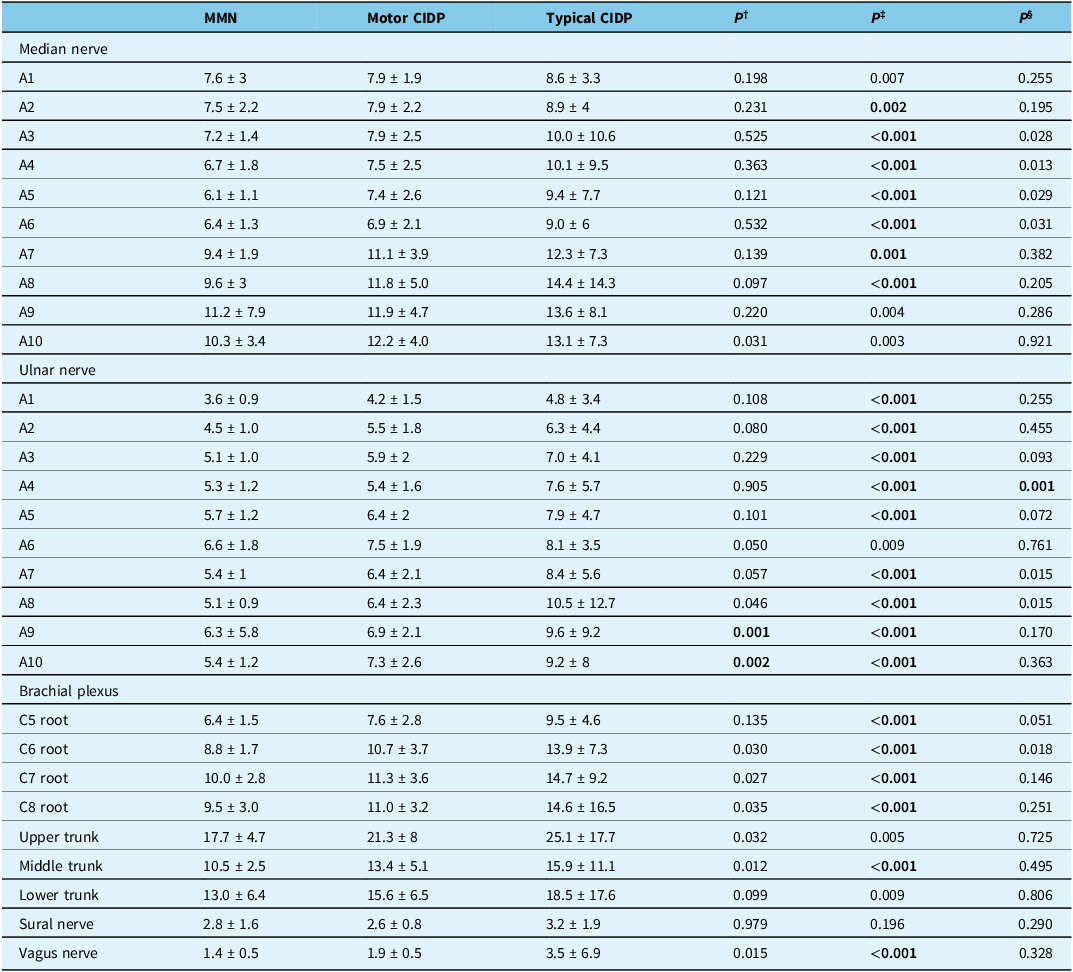
Table 2 shows the CSA of the median nerve, ulnar nerve, brachial plexus, sural nerve and vagus nerve in the MMN, motor CIDP and typical CIDP groups. CSAs at nearly all sites were significantly larger in patients with typical CIDP than in those with MMN. CSAs were significantly larger in patients with motor CIDP than in those with MMN at some proximal sites of the ulnar nerves (A9–10 of the ulnar nerve) Supplemental Material 1 shows the mean and dispersion of the CSA at each site on the nerves in the MMN, motor CIDP and typical CIDP groups. The dispersion of CSAs in typical CIDP patients was significantly greater than that in MMN and motor CIDP patients. Figure 1A–C shows the mean CSA at each site in the MMN, PM-CIDP, MPred-CIDP and typical CIDP groups. The CSAs in PM-CIDP patients were close to the MMN, while those in MPred-CIDP patients were close to the typical CIDP patients. The CSAs of MMN, PM-CIDP, MPred-CIDP and typical CIDP gradually formed a continuum.
Cross-sectional area (CSA) in multiple motor neuropathy (MMN), pure motor chronic inflammatory demyelinating polyneuropathy (CIDP), motor predominant CIDP and typical CIDP. CSA (mean ± SD, mm2) at different sites on the median nerve (A), ulnar nerve (B) and brachial plexus (C) in the MMN, pure motor CIDP (PM-CIDP), motor predominant CIDP (MPred-CIDP) and typical CIDP groups. To demonstrate the comparison of the mean CSA more adequately, some error bars were outside of the range.

Figure 1 Long description
Three line graphs compare cross-sectional area (CSA) in different types of neuropathy across median nerve, ulnar nerve, and brachial plexus. The first graph (A) shows CSA measurements at different sites on the median nerve for multiple motor neuropathy (MMN), pure motor chronic inflammatory demyelinating polyneuropathy (CIDP), motor predominant CIDP, and typical CIDP. The second graph (B) displays similar measurements for the ulnar nerve. The third graph (C) presents normalized CSA values for the brachial plexus. Each graph includes error bars to indicate variability, with some error bars extending beyond the range for better comparison. The data points are color-coded: green for typical CIDP, red for motor predominant CIDP, purple for pure motor CIDP, and blue for MMN. The x-axes represent different measurement sites, while the y-axes represent CSA in square millimeters. The graphs illustrate the differences in CSA among the various types of neuropathy.
Cross-sectional area (mean ± SD, mm2) at different sites of the median and ulnar nerves and brachial plexus in MMN patients, motor CIDP patients and typical CIDP patients

Table 2 Long description
The table presents cross-sectional area (CSA) measurements in square millimeters for the median nerve, ulnar nerve, brachial plexus, sural nerve, and vagus nerve across three groups: MMN, motor CIDP, and typical CIDP. The table has 10 rows for the median nerve, 10 rows for the ulnar nerve, and 8 rows for the brachial plexus, with each row representing different sites (A1 to A10 for nerves, and specific roots and trunks for the brachial plexus). Each column represents a different group: MMN, motor CIDP, and typical CIDP, along with p-values for statistical comparisons. Notable trends include significantly larger CSAs in typical CIDP patients compared to MMN patients at nearly all sites. Additionally, motor CIDP patients show significantly larger CSAs than MMN patients at specific proximal sites of the ulnar nerve (A9 and A10). The dispersion of CSAs in typical CIDP patients is significantly greater than in MMN and motor CIDP patients. The data highlights a continuum of CSA measurements from MMN to typical CIDP, with PM-CIDP patients showing CSAs close to MMN and MPred-CIDP patients showing CSAs close to typical CIDP.
Note: CSA = cross-sectional area; CIDP = chronic inflammatory demyelinating polyradiculoneuropathy; MMN = multiple motor neuropathy; SD = standard deviation.
†Statistical comparison between MMN and motor CIDP (Mann–Whitney U test).
‡Statistical comparison between MMN and typical CIDP (Mann–Whitney U test).
§Statistical comparison between motor CIDP and typical CIDP (Mann–Whitney U test). According to the Holm–Bonferroni correction, P ≤ 0.002 was considered significant for the median nerve, P ≤ 0.002 was considered significant for the ulnar nerve and P < 0.001 was considered significant for the brachial plexus (more details in Supplemental Material 3).
Statistically significant P values are bold.
The extent of enlargement in the forearm segment, arm segment and brachial plexus of the MMN, motor CIDP and typical CIDP was shown in Figure 2. For MMN and typical CIDP, the ratio of no enlargement to enlargement was similar among the forearm segment, arm segment and brachial plexus. For motor CIDP, the percentage of mild and significant enlargement was larger in the arm segment compared with the forearm segment and was larger in the brachial plexus compared with the arm segment.
Extent of enlargement in multiple motor neuropathy (MMN), motor chronic inflammatory demyelinating polyneuropathy (CIDP) and typical CIDP. The extent of enlargement was shown in the forearm segment, arm segment and Erb’s point on the MMN, motor CIDP and CIDP, respectively.

Figure 2 Long description
The bar graph compares the extent of enlargement in multiple motor neuropathy (MMN), motor chronic inflammatory demyelinating polyneuropathy (CIDP), and typical CIDP across three segments: forearm segment, arm segment, and Erb's point. The x-axis labels the conditions (MMN, motor CIDP, CIDP) for each segment, while the y-axis represents the percentage of enlargement. The graph uses a stacked bar format with three colors: blue for no enlargement, orange for mild enlargement, and gray for significant enlargement. In the forearm segment, MMN shows 57.1% no enlargement, 31% mild enlargement, and 11.9% significant enlargement. Motor CIDP shows 41.4% no enlargement, 27.6% mild enlargement, and 31% significant enlargement. CIDP shows 20.6% no enlargement, 36.4% mild enlargement, and 43% significant enlargement. In the arm segment, MMN shows 57.1% no enlargement, 25.7% mild enlargement, and 17.1% significant enlargement. Motor CIDP shows 34.5% no enlargement, 41.4% mild enlargement, and 24.1% significant enlargement. CIDP shows 23.2% no enlargement, 31.1% mild enlargement, and 45.6% significant enlargement. In Erb's point, MMN shows 54.3% no enlargement, 37.1% mild enlargement, and 8.6% significant enlargement. Motor CIDP shows 25.9% no enlargement, 40.7% mild enlargement, and 33.3% significant enlargement. CIDP shows 22% no enlargement, 40.1% mild enlargement, and 37.9% significant enlargement. All values are approximated.
Figure 3 shows the ultrasonography of the median nerve (A4 and A8) and cervical roots (C6 and C7) in an MMN patient, pure motor CIDP patient and CIDP patient.
Ultrasonography in multiple motor neuropathy (MMN), motor chronic inflammatory demyelinating polyneuropathy (CIDP) and CIDP patients. Ultrasonography of the left median nerve (A4 and A8 site) and left cervical roots (C6 and C7 roots) of MMN (first row), pure motor CIDP (second row) and typical CIDP (third row) patients.

Figure 3 Long description
The image shows a series of ultrasound scans of the left median nerve and left cervical roots for patients with multifocal motor neuropathy (MMN), pure motor chronic inflammatory demyelinating polyneuropathy (CIDP), and typical CIDP. The scans are organized in three rows, each representing a different condition. The first row shows the scans for an MMN patient, the second row for a pure motor CIDP patient, and the third row for a typical CIDP patient. Each row contains four images, labeled with specific sites: A4 and A8 for the median nerve, and C6 and C7 for the cervical roots. The scans highlight the cross-sectional areas and intranerve variability, which are key features in differentiating these conditions. The labels and measurements provide detailed information about the nerve structures and their abnormalities.
The intranerve CSA variability of the median nerve was 2.4 ± 1.6, 2.1 ± 0.7 and 2.7 ± 2.7 for MMN, motor CIDP and typical CIDP, respectively. The intranerve CSA variability of the ulnar nerve was 2.5 ± 1.9, 2.2 ± 0.6 and 3.2 ± 3.1 for MMN, motor CIDP and typical CIDP, respectively. The intranerve CSA variability of the ulnar nerve was greater in typical CIDP patients than in MMN patients. The internerve CSA variability was 1.83 ± 0.67 in MMN, 1.49 ± 0.31 in motor CIDP and 1.87 ± 0.96 in typical CIDP. The internerve CSA variability was lower in motor CIDP patients than in typical CIDP or MMN patients without statistical significance.
Nerve conduction studies
The results of the motor NCSs of MMN, motor CIDP and typical CIDP are shown in Table 3. The DML was significantly more delayed in typical CIDP than in MMN or motor CIDP. MCV was significantly lower in typical CIDP than in MMN or motor CIDP. The CMAPerb/CMAPaxilla of the median nerve was significantly greater in the MMN group than in the motor CIDP or typical CIDP group. CB or probable CB was observed in 63.2% (12/19) MMN patients and 60% (9/15) motor CIDP patients, compared to 44.4% (52/117) typical CIDP patients. The results of the motor NCSs of MMN, PM-CIDP, MPred-CIDP and typical CIDP are shown in Supplemental Material 2. The MCV of the median and ulnar nerves was high in PM-CIDP patients, close to the MMN, and was low in MPred-CIDP patients, close to the typical CIDP patients.
Nerve conduction studies in patients with MMN, motor CIDP and typical CIDP

Table 3 Long description
The table presents a comparison of nerve conduction studies across three groups: MMN, motor CIDP, and typical CIDP. It includes measurements such as CMAP wrist, CMAP erb, DML, MCV elbow-wrist, MCV axilla-elbow, CMAP erb/CMAP axilla, and TLI for both median and ulnar nerves. The table has 18 rows and 8 columns, with columns labeled MMN, Motor CIDP, Typical CIDP, and P values for different comparisons. Notable trends include significant differences in DML and MCV values among the groups, with typical CIDP showing more delayed DML and lower MCV. The CMAP erb/CMAP axilla ratio for the median nerve is significantly higher in the MMN group. Conduction block percentages are also compared across the groups.
Note: CIDP = chronic inflammatory demyelinating polyneuropathy; MMN = multiple motor neuropathy; SD = standard deviation; CMAP = compound motor action potential; DML = distal motor latency; MCV = motor conduction velocity; TLI = terminal latency index; CB = conduction block.
TLI = terminal distance (55 mm)/(distal motor latency (ms)×MCV1 (m/s)).
†Statistical comparison between MMN and motor CIDP (Mann–Whitney U test).
‡Statistical comparison between MMN and typical CIDP (Mann–Whitney U test).
§Statistical comparison between motor CIDP and typical CIDP (Mann–Whitney U test).
According to the Bonferroni correction, P < 0.017 was considered significant.
The chi-square test was used for comparisons of CB.
Nerve ultrasound and nerve conduction studies for the differential diagnosis of MMN from motor CIDP
ROC curves for distinguishing MMN from motor CIDP were performed to assess the diagnostic value of the CSA of the median nerve at A10 site, maximum CSA of the ulnar nerve at A8–10 sites, maximal CSA of the C6–8 roots, CMAPerb/CMAPaxilla of the median nerve, CMAPerb/CMAPaxilla of the ulnar nerve, CMAPerb of the ulnar nerve, MCVbl elbow-wrist of the ulnar nerve, MCVab-bl elbow of the ulnar nerve and MCVaxilla-ab elbow of the ulnar nerve (Figure 4A–D). The AUCs were above 0.700 for the maximum CSA of the ulnar nerve at sites A8–10, CMAPerb/CMAPaxilla of the median nerve, CMAPerb of the ulnar nerve and MCVab-bl elbow of the ulnar nerve. The combination of the maximum CSA of the ulnar nerve at sites A8–10 and the CMAPerb of the ulnar nerve had an AUC of 0.773.
Receiver operating characteristic (ROC) curves for distinguishing multiple motor neuropathy (MMN) and motor chronic inflammatory demyelinating polyneuropathy (CIDP). ROC curves for cross-sectional area (CSA) of median nerve A10 site, maximum CSA of the ulnar nerve A8–10 sites, maximal CSA of C6–8 roots, CMAPerb/CMAPaxilla of the median nerve, CMAPerb/CMAPaxilla of the ulnar nerve, CMAPerb of the ulnar nerve, MCVbl elbow-wrist of the ulnar nerve, MCVab-bl elbow of the ulnar nerve and MCVaxilla-ab elbow of the ulnar nerve for distinguishing MMN and motor CIDP. (A) CSA of the median nerve A10 site, maximum CSA of the ulnar nerve A8–10 site and maximal CSA of C6–8 roots. (B) CMAPerb/CMAPaxilla of the median nerve, CMAPerb/CMAPaxilla of the ulnar nerve and CMAPerb of the ulnar nerve. (C) MCVbl elbow-wrist of the ulnar nerve, MCVab-bl elbow of the ulnar nerve and MCVaxilla-ab elbow of the ulnar nerve. (D) Combination of the maximum CSA at the ulnar A8–10 site and the CMAPerb of the ulnar nerve.

Figure 4 Long description
Four line graphs showing ROC curves for distinguishing multiple motor neuropathy (MMN) from motor chronic inflammatory demyelinating polyneuropathy (CIDP) using various nerve measurements. The x-axis represents one minus specificity, and the y-axis represents sensitivity. Graph A shows the ROC curves for the cross-sectional area (CSA) of the median nerve at the A10 site, the maximum CSA of the ulnar nerve at A810 sites, and the maximal CSA of the C68 roots. Graph B shows the ROC curves for the CMAPerb/CMAPaxilla of the median nerve, the CMAPerb/CMAPaxilla of the ulnar nerve, and the CMAPerb of the ulnar nerve. Graph C shows the ROC curves for the MCVbl elbow-wrist of the ulnar nerve, the MCVab-bl elbow of the ulnar nerve, and the MCVaxilla-ab elbow of the ulnar nerve. Graph D shows the ROC curve for the combination of the maximum CSA at the ulnar A810 site and the CMAPerb of the ulnar nerve. All values are approximated.
Discussion
Both MMN and motor CIDP are chronic progressive immune-mediated peripheral neuropathies without sensory loss. The underlying disease mechanism, however, is considered to be different. MMN is hypothesized to be an axonopathy without overt segmental demyelination or onion bulb formation, and antibody-mediated attack directed against components of axolemma at nodes of Ranvier caused CB, transitory paranodal demyelination and remyelination and axonal degeneration and regeneration. Reference Taylor, Dyck, Engelstad, Gruener, Grant and Dyck22,Reference Yeh, Dyck, van den Berg, Kiernan and Taylor23 Motor CIDP, on the other hand, is a subtype of CIDP, which is a spectrum of demyelinating neuropathies with cell-mediated and humoral mechanisms that act together. Reference Mathey, Park and Hughes24 However, previous studies have shown similarities in NCS and treatment responses between MMN and motor CIDP. MCV reduction was less frequent, and CB was more frequent in both disorders. Reference Pegat, Boisseau and Maisonobe3–Reference Li, Niu and Liu8 Treatment with prednisone is generally ineffective in treating MMN and a great part of motor CIDP. Reference Van den Bergh, van Doorn and Hadden1,Reference Van Asseldonk, Franssen, Van den Berg-Vos, Wokke and Van den Berg2,Reference Kimura, Sakurai and Koumura5 We investigated the similarities and differences among MMN, motor CIDP and typical CIDP through conduction studies and nerve ultrasound. Electrophysiological studies showed that, compared with those in typical CIDP patients, MCV reduction and DML elongation were less frequent in both MMN and motor CIDP patients, while CB occurred more frequently in both MMN and motor CIDP patients. Compared with motor CIDP, MMN had a greater CMAP at Erb’s point, indicating less proximal involvement in MMN. Further investigation showed that the MCV in PM-CIDP patients was faster, close to MMN, while the MCV in MPred-CIDP patients was slower, close to typical CIDP. Ultrasonography revealed that the CSAs in PM-CIDP patients were more close to MMN, while those in MPred-CIDP patients were more close to typical CIDP patients. The CSAs of MMN, PM-CIDP, MPred-CIDP and typical CIDP formed a continuum. The electrophysiological and morphological performance of MMN, PM-CIDP, MPred-CIDP and typical CIDP might constitute a spectrum.
In our cohort, 9/15 motor CIDP patients were treated with steroids. Among them, half of the PM-CIDP patients (3/6) and all the MPred-CIDP patients (3/3) were responsive. In contrast, 93.4% of typical CIDP patients and none of the MMN patients (3/3) were steroid responsive. In support of our findings, previous studies have shown a low response rate to steroids in PM-CIDP patients (0%–50%) and a high response rate in MPred-CIDP patients (100%). Reference Pegat, Boisseau and Maisonobe3–Reference Sabatelli, Madia, Mignogna, Lippi, Quaranta and Tonali6,Reference Busby and Donaghy25 The treatment response to steroids in PM-CIDP was similar to that in MMN, while that in MPred-CIDP was similar to that in typical CIDP. For patients with MPred-CIDP, steroids might be considered.
Nerve ultrasound revealed that the percentage of mild and significant enlargements was higher in the proximal segment than in the distal segment in motor CIDP patients. At nearly all sites, the CSAs were smaller in the MMN than in the typical CIDP. The CSAs in PM-CIDP patients were close to the MMN, while those in MPred-CIDP patients were close to the typical CIDP patients. The CSAs of MMN, PM-CIDP, MPred-CIDP and typical CIDP formed a continuum. Previous studies have shown less ultrasonographic enlargement in MMN than in CIDP. Merola et al. reviewed peripheral nerve ultrasound findings in CIDP and MMN patients and reported greater side-to-side intranerve variability in MMN and greater CSA in CIDP patients than in HCs. Reference Merola, Rosso, Romagnolo, Peci and Cocito9 Kerasnoudis et al. used the Bochum ultrasound score (BUS) to distinguish CIDP from MMN and multifocal acquired demyelinating sensory and motor neuropathy (MADSAM). A cutoff value of ≥2 points on the BUS showed a sensitivity of 80% and specificity of 87.5% in distinguishing CIDP from MMN or MADSAM. Reference Kerasnoudis, Pitarokoili, Gold and Yoon10 However, there has been no research on the ultrasonographic characteristics of motor CIDP. The proximal enlargement in motor CIDP might reflect its distinct underlying pathological changes. The continuum condition formed by the CSA of the MMN, PM-CIDP, MPred-CIDP and typical CIDP might be an indication of the underlying pathological changes in these disorders.
A comparison of the electrophysiological features among the MMN, motor CIDP and typical CIDP groups showed greater similarity between MMN and motor CIDP. The DML was significantly longer in typical CIDP than in MMN or motor CIDP. MCV was significantly lower in typical CIDP than in MMN or motor CIDP. There was more CB in the MMN or motor CIDP groups than in the typical CIDP group. Supporting our findings, most previous studies have shown a high percentage of CB and a low percentage of reduced CV in motor CIDP patients. Paget et al. reported that CB occurred in 82% (14/17) of motor CIDP patients, while a reduced CV occurred in only 35% (6/17) of patients. Reference Pegat, Boisseau and Maisonobe3 Doneddu et al. reported that 65% (11/17) of pure motor patients had CB, and 53% (9/17) of patients had reduced MCV. Reference Doneddu, Cocito and Manganelli4 Kimura et al. reported that 100% (5/5) of patients had reduced MCV, 60% (3/5) had prolonged distal motor latencies and 80% (4/5) had CB in motor CIDP. Reference Kimura, Sakurai and Koumura5 Sabatelli et al. showed that CB occurred in 100% (4/4) of pure motor CIDP patients. Reference Sabatelli, Madia, Mignogna, Lippi, Quaranta and Tonali6 Zhou et al. compared nerve conduction parameters between MMN and CIDP patients and reported that the MCV was lower in CIDP patients with CB than in MMN patients. Reference Zhou, Zhu and Zhu7 We further compared the MMN and motor CIDP and between subtypes of motor CIDP. A comparison of the MMN with the motor CIDP showed that the CMAPerb/CMAPaxilla ratio and CMAPerb were greater in the MMN than in the motor CIDP. This finding implicates more proximal involvement in motor CIDP, in accordance with the ultrasonographic results. MCV was fast in PM-CIDP patients, close to the MMN, and MCV was slow in MPred-CIDP patients, close to the typical CIDP. The MCV of MMN, PM-CIDP, MPred-CIDP and typical CIDP also formed a continuum.
Our work has several limitations. First, not all patients were treatment naïve. Some patients were treated with steroids or IVIg prior to enrollment. The effect of treatment on CSA in CIDP patients has been discussed in previous work. Reference Niu, Li and Liu15 Second, the disease duration differed among the groups. Disease duration might have an impact on CSA in some CIDP patients, as CSA increases during follow-up in some CIDP patients. Reference Niu, Li and Liu15 In the MMN, a follow-up study by Rattay et al. showed that the ultrasound pattern sum score was not correlated with disease duration. Reference Rattay, Winter and Decard26 Third, the number of patients with PM-CIDP and MPred-CIDP was too small to further statistically compare them with MMN or typical CIDP. Fourth, it would be more compelling if nerve pathological studies were also included to demonstrate that MMN, PM-CIDP, MPred-CIDP and typical CIDP are the spectrum of disorders. Last, the ultrasound examiner was unblinded to the patient diagnosis, which might be a potential source of bias.
In summary, we have described the comparison of ultrasonographic and electrophysiological features among MMN patients, motor CIDP patients and typical CIDP patients. Both nerve ultrasound and NCS showed more prominent proximal involvement in motor CIDP than in MMN. The CSA of the MMN, pure motor CIDP, motor predominant CIDP and typical CIDP formed a continuum, and so did MCV. This study provides a new perspective for examining the pathological mechanisms of these chronic immune peripheral neuropathies.
Supplementary material
The supplementary material for this article can be found at https://doi.org/10.1017/cjn.2025.10136.
Acknowledgments
None.
Author contributions
JWN: clinical studies, data acquisition and analysis, statistical analysis, manuscript preparation; QYD: data acquisition; NH: statistical analysis; LYC: concept; MSL: concept, design, manuscript editing and review.
Funding statement
This study was funded by the National High Level Hospital Clinical Research Funding (2022-PUMCH-B-017, 2022-PUMCH-D-002) and CAMS Innovation Fund for Medical Sciences (CIFMS 2021-I2M-1-003).
Competing interests
None.






Target article
Nerve Ultrasound of Multifocal Motor Neuropathy and Motor and Typical Chronic Inflammatory Demyelinating Neuropathy
Related commentaries (1)
Reviewer Comment on Niu et al. “Nerve Ultrasound of Multifocal Motor Neuropathy, Motor and Typical Chronic Inflammatory Demyelinating Neuropathy”