






No CrossRef data available.
Article contents
Beyond a gut feeling: How the immune system impacts the effect of gut microbiota in neurodevelopment
Published online by Cambridge University Press: 15 July 2019
Abstract
Hooks et al. posit that gastrointestinal microbes alter the end state of development indirectly. Here, we present the immune system as the link that facilitates communication between the gut and the brain. Illustrating the case of autism spectrum disorder, we explicate the role of the immune system in responding to microbial dysbiosis by inducing an inflammatory state that affects neurodevelopment. We propose two models: directly, within the infant, and indirectly, via maternal and infant systems.
Information
- Type
- Open Peer Commentary
- Information
- Copyright
- Copyright © Cambridge University Press 2019
References
American Psychiatric Association (2013) Diagnostic and statistical manual of mental disorders (DSM-5). American Psychiatric Association.Google Scholar
Ashwood, P. & Wakefield, A. J. (2006) Immune activation of peripheral blood and mucosal CD3+ lymphocyte cytokine profiles in children with autism and gastrointestinal symptoms. Journal of Neuroimmunology 173(1–2):126–34.Google Scholar
Azhari, A., Farouq, M. & Esposito, G. (2018) A systematic review of gut-immune-brain mechanisms in autism spectrum disorder. Developmental Psychobiology. Available at: https://doi.org/10.1002/dev.21803.Google Scholar
Breese, E. J., Michie, C. A., Nicholls, S. W., Murch, S. H., Williams, C. B., Domizio, P., Walker-Smith, J. A. & Macdonald, T. T. (1994) Tumor necrosis factor α-producing cells in the intestinal mucosa of children with inflammatory bowel disease. Gastroenterology 106(6):1455–66.Google Scholar
Carabotti, M., Scirocco, A., Maselli, M. A. & Severi, C. (2015) The gut-brain axis: Interactions between enteric microbiota, central and enteric nervous systems. Annales de Gastroenterologie et D'hepatologie 28(2):203–209.Google Scholar
El-Ansary, A. & Al-Ayadhi, L. (2014) GABAergic/glutamatergic imbalance relative to excessive neuroinflammation in autism spectrum disorders. Journal of Neuroinflammation 11:189.Google Scholar
Erny, D., Hrabě de Angelis, A. L., Jaitin, D., Wieghofer, P., Staszewski, O., David, E., Keren-Shaul, H., Mahlakoiv, T., Jakobshagen, K., Buch, T., Schwierzeck, V., Utermöhlen, O., Chun, E., Garrett, W. S., McCoy, K. D., Diefenbach, A., Staeheli, P., Stecher, B., Amit, I. & Prinz, M. (2015) Host microbiota constantly control maturation and function of microglia in the CNS. Nature Neuroscience 18(7):965–77.Google Scholar
Fernández de Cossío, L., Guzmán, A., van der Veldt, S. & Luheshi, G.N. (2017) Prenatal infection leads to ASD-like behavior and altered synaptic pruning in the mouse offspring. Brain, Behavior, and Immunity 63:88–98.Google Scholar
Finegold, S. M., Molitoris, D., Song, Y., Liu, C., Vaisanen, M.-L., Bolte, E., McTeague, M., Sandler, R., Wexler, H., Marlowe, E. M., Collins, M. D., Lawson, P. A., Summanen, P., Baysallar, M., Tomzynski, T. J., Read, E., Johnson, E., Rolfe, R., Nasir, P., Shah, H., Haake, D. A., Manning, P. & Kaul, A. (2002) Gastrointestinal microflora studies in late-onset autism. Clinical Infectious Diseases 35(S1):S6–16.Google Scholar
Gilmore, J. H., Jarskog, L. F. & Vadlamudi, S. (2005) Maternal poly I:C exposure during pregnancy regulates TNF alpha, BDNF, and NGF expression in neonatal brain and the maternal-fetal unit of the rat. Journal of Neuroimmunology 159(1–2):106–12.Google Scholar
Kang, D.-W., Park, J. G., Ilhan, Z. E., Wallstrom, G., Labaer, J., Adams, J. B. & Krajmalnik-Brown, R. (2013) Reduced incidence of Prevotella and other fermenters in intestinal microflora of autistic children. PLoS ONE 8(7):e68322.Google Scholar
Khakzad, M. R., Javanbakht, M., Shayegan, M. R., Kianoush, S., Omid, F., Hojati, M. & Meshkat, M. (2012) The complementary role of high sensitivity C-reactive protein in the diagnosis and severity assessment of autism. Research in Autism Spectrum Disorders 6(3):1032–37.Google Scholar
Kim, S., Kim, H., Yim, Y. S., Ha, S., Atarashi, K., Tan, T. G., Longman, R. S., Honda, K., Littman, D. R., Choi, G. B. & Huh, J. R. (2017b) Maternal gut bacteria promote neurodevelopmental abnormalities in mouse offspring. Nature 549(7673):528–32.Google Scholar
Li, X., Chauhan, A., Sheikh, A. M., Patil, S., Chauhan, V., Li, X.-M., Ji, L., Brown, T. & Malik, M. (2009) Elevated immune response in the brain of autistic patients. Journal of Neuroimmunology 207(1–2):111–16.Google Scholar
Qin, L., Wu, X., Block, M. L., Liu, Y., Breese, G. R., Hong, J.-S., Knapp, D. J. & Crews, F. T. (2007) Systemic LPS causes chronic neuroinflammation and progressive neurodegeneration. GLIA 55(5):453–62.Google Scholar
Rodriguez, J. I. & Kern, J. K. (2011) Evidence of microglial activation in autism and its possible role in brain underconnectivity. Neuron Glia Biology 7(2–4):205–13.Google Scholar
Takano, T. (2015) Role of microglia in autism: Recent advances. Developmental Neuroscience 37(3):195–202.Google Scholar
Tetreault, N. A., Hakeem, A. Y., Jiang, S., Williams, B. A., Allman, E., Wold, B. J. & Allman, J. M. (2012) Microglia in the cerebral cortex in autism. Journal of Autism and Developmental Disorders 42(12):2569–84.Google Scholar
Torrente, F., Ashwood, P., Day, R., Machado, N., Furlano, R. I., Anthony, A., Davies, S. E., Wakefield, A. J., Thomson, M. A., Walker-Smith, J. A. & Murch, S. H. (2002) Small intestinal enteropathy with epithelial IgG and complement deposition in children with regressive autism. Molecular Psychiatry 7:375–82.Google Scholar
Vargas, D. L., Nascimbene, C., Krishnan, C., Zimmerman, A. W. & Pardo, C. A. (2004) Neuroglial activation and neuroinflammation in the brain of patients with autism. Annals of Neurology 57(1):67–81.Google Scholar
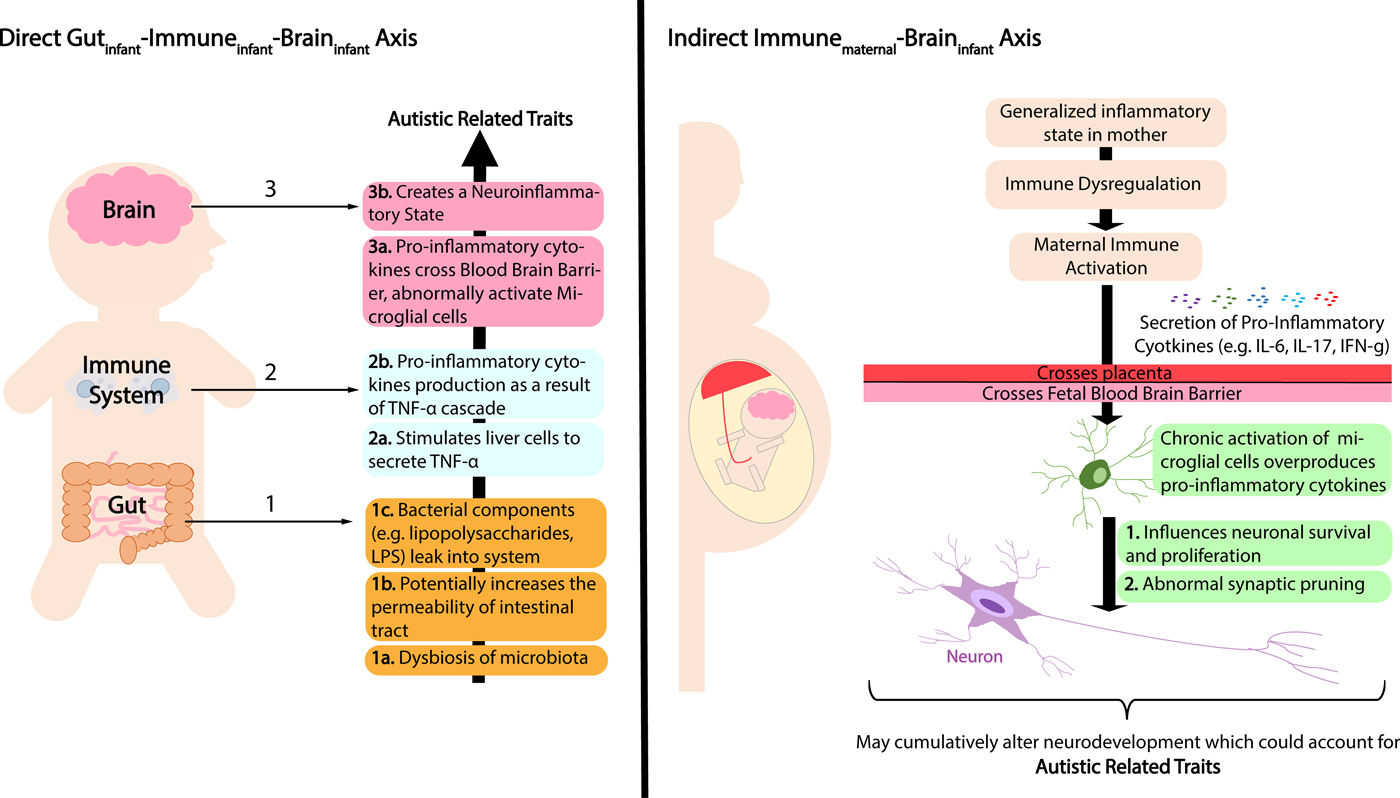
Figure 1. Schematic diagram illustrating theoretical models of direct (gutinfant-immuneinfant-braininfant) and indirect (immunematernal-braininfant) mechanisms underlying autism spectrum disorder (ASD). The direct model features atypicality in the infant's gastrointestinal microbiota, leading to system-wide neuroinflammation in the infant. The indirect model features dysregulated maternal gastrointestinal microbiota, instigating maternal immune activation (MIA) and subsequent neuroinflammation in the infant. Neuroinflammation stemming from either of the two mechanisms will compromise neurodevelopment and induce the emergence of autistic-like traits.
Target article
Related commentaries (19)
A call for mapping the development of the microbiota-gut-brain axis during human infancy
Beyond a gut feeling: How the immune system impacts the effect of gut microbiota in neurodevelopment
Nourishing the gut microbiota: The potential of prebiotics in microbiota-gut-brain axis research
Practical guidelines for gut microbiome analysis in microbiota-gut-brain axis research
The parent-offspring microbiome and neurobehavioral development
The gut microbiota has been recurrently reported to influence developmental outcomes (e.g., Finegold et al. Reference Finegold, Molitoris, Song, Liu, Vaisanen, Bolte, McTeague, Sandler, Wexler, Marlowe, Collins, Lawson, Summanen, Baysallar, Tomzynski, Read, Johnson, Rolfe, Nasir, Shah, Haake, Manning and Kaul2002; Kang et al. Reference Kang, Park, Ilhan, Wallstrom, Labaer, Adams and Krajmalnik-Brown2013). Hooks et al. proposed that the mere presence of microbes in the gut triggers a response in the developing organism that results in an altered end state of development, rather than the microbes being a direct causative agent. Although we concur with this point, it is important to further address it in relation to the immune system and how pivotal it is as an intermediary system that affects neurodevelopment (Fig. 1). To illustrate our point, we present the case of autism spectrum disorder (ASD), a neurodevelopmental condition that features restricted behaviors and deficits in social communication (American Psychiatric Association 2013).
Schematic diagram illustrating theoretical models of direct (gutinfant-immuneinfant-braininfant) and indirect (immunematernal-braininfant) mechanisms underlying autism spectrum disorder (ASD). The direct model features atypicality in the infant's gastrointestinal microbiota, leading to system-wide neuroinflammation in the infant. The indirect model features dysregulated maternal gastrointestinal microbiota, instigating maternal immune activation (MIA) and subsequent neuroinflammation in the infant. Neuroinflammation stemming from either of the two mechanisms will compromise neurodevelopment and induce the emergence of autistic-like traits.
The comorbidity of ASD and gastrointestinal disorders has been often reported (e.g., Ashwood & Wakefield Reference Ashwood and Wakefield2006; Torrente et al. Reference Torrente, Ashwood, Day, Machado, Furlano, Anthony, Davies, Wakefield, Thomson, Walker-Smith and Murch2002). In our recent review, we showed that the risk of ASD increases with respect to the inflammatory state, rather than to the presence of a specific species of microbe, underscoring how system-wide changes in inflammatory profiles may direct neurodevelopmental trajectories (Azhari et al. Reference Azhari, Farouq and Esposito2018). Indeed, the response to gut microbiota that the organism launches is primarily composed of signals from immuno-inflammation pathways, allowing for a relay between the gut and the central nervous system (CNS) (Carabotti et al. Reference Carabotti, Scirocco, Maselli and Severi2015; Erny et al. Reference Erny, Hrabě de Angelis, Jaitin, Wieghofer, Staszewski, David, Keren-Shaul, Mahlakoiv, Jakobshagen, Buch, Schwierzeck, Utermöhlen, Chun, Garrett, McCoy, Diefenbach, Staeheli, Stecher, Amit and Prinz2015). This gut-immune-brain communication can be traced to several main points of contact across the systems, primarily facilitated by signaling immune molecules. The gastrointestinal microbiota has significant influence over the profile of certain circulating pro-inflammatory cytokines, chemokines, and growth factors, such as IFN-γ (interferon-γ), IL-17 (interleukin-17), IL-6 (interleukin-6), and TNF-α (tumor necrosis factor-α) (e.g., El-Ansary & Al-Ayadhi Reference El-Ansary and Al-Ayadhi2014). The link between autism and immune dysfunction has been asserted by findings of atypical upregulation of these cytokines in persons with autism (e.g., Khakzad et al. Reference Khakzad, Javanbakht, Shayegan, Kianoush, Omid, Hojati and Meshkat2012; Li et al. Reference Li, Chauhan, Sheikh, Patil, Chauhan, Li, Ji, Brown and Malik2009).
Pro-inflammatory cytokines function as signaling molecules and have the capacity to communicate with the CNS, serving as a bridge between the gut microbiota and the brain. Indeed, post-mortem studies on brain tissues obtained from deceased persons with autism showed the presence of enhanced neuroinflammation in several brain regions, including the cerebral cortex, white matter, and cerebellum (e.g., Vargas et al. Reference Vargas, Nascimbene, Krishnan, Zimmerman and Pardo2004). Pro-inflammatory molecules responsible for this have been postulated to impede brain development indirectly, through non-neural cells. In individuals with ASD, a type of resident non-neural cell that has been found to be activated at atypical levels constitutes the microglia (e.g., Tetreault et al. Reference Tetreault, Hakeem, Jiang, Williams, Allman, Wold and Allman2012). Although moderate activation of the microglia in response to injury or infection in the CNS is protective, chronic microglial activation compromises brain development in one of two ways. First, abnormally activated microglia overproduce pro-inflammatory cytokines, which contributes to damage in synaptic networks and neuronal cell death (Rodriguez & Kern Reference Rodriguez and Kern2011). Second, chronically activated microglia are responsible for elevated occurrences of phagocytosis and excessive removal of neuronal debris, leading to impaired neural development (Takano Reference Takano2015). Dysfunctional microglia are no longer sensitive to external immune signals either, which leads to a perpetuation of dysregulated phagocytosis (Fernández de Cossío et al. Reference Fernández de Cossío, Guzmán, van der Veldt and Luheshi2017). At present, accumulating evidence drives at one possible hypothesis: In autistic persons, elevated levels of pro-inflammatory cytokines may stem from the instigation of the gut microbiota onto the immune system; these immune signals contribute to neuroinflammation that ultimately hinders neurodevelopment. This theory posits a gutinfant-immuneinfant-braininfant model, where biological pathways from the gut microbiota to the eventual emergence of the autistic phenotype occur within an individual. Although this hypothesis is intriguing, at present, researchers have yet to elucidate a causal pathway that proves that the dysregulation of pro-inflammatory cytokines attributable to dysbiosis of the gut microbiota is the same immune phenomenon that leads to chronically activated microglial cells in the brain of ASD individuals.
As neurodevelopment begins early in the course of fetal maturation, it is important that we address the mechanism at the prenatal phase too. Hooks et al. refuted the postulation of the gut microbiota exerting any neurodevelopmental impact given that the in utero environment is sterile. Although the notion of in utero sterility may be true, the immune system of the mother, however, could still impact the fetus during gestation. We posit that the state of generalized inflammation in the pregnant mother, maternal immune activation (MIA), potentially alters neurodevelopment in infants (e.g., Gilmore et al. Reference Gilmore, Jarskog and Vadlamudi2005). This theory has largely been supported by animal studies, such as that conducted by Kim et al. (Reference Kim, Kim, Yim, Ha, Atarashi, Tan, Longman, Honda, Littman, Choi and Huh2017b). In this mice study, the authors showed that specific maternal gut microbes are associated with an increase in pro-inflammatory IL-17 in the mother, along with the appearance of autistic behaviors in the offspring. A study on ASD patients showed that TNF-α, potentially secreted by liver cells in the presence of gut lipopolysaccharides (LPS), creates a peripheral inflammation that results in microglia activation in the brain (e.g., Breese et al. Reference Breese, Michie, Nicholls, Murch, Williams, Domizio, Walker-Smith and Macdonald1994; Qin et al. Reference Qin, Wu, Block, Liu, Breese, Hong, Knapp and Crews2007). As opposed to the first model, this second model presents an indirect immunematernal-braininfant pathway that involves cross-talk between the immune system of the mother and the CNS of the infant.
In conclusion, we have presented the case of ASD as a neurodevelopmental condition involving both the gut microbiota and the immune system. We have also proposed two theoretical models that the field should consider (Azhari et al. Reference Azhari, Farouq and Esposito2018). The first model features direct gut-immune-brain association within the individual at the postnatal phase, whereas the second model is an indirect model implicating both maternal and infant systems in the prenatal phase. These theoretical models may sprout experimental paradigms that allow for existing postulations to be tested and, in doing so, uncover causal pathways from the gut to the immune system and, ultimately, to the brain.